. 2010 Feb 9;107(6):2705-10.
doi: 10.1073/pnas.0915001107. Epub 2010 Jan 25.

Bimodular auxin response controls organogenesis in Arabidopsis

Affiliations

Bimodular auxin response controls organogenesis in Arabidopsis

Ive De Smet et al. Proc Natl Acad Sci U S A. .

Abstract

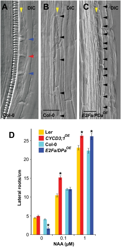
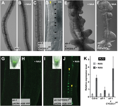
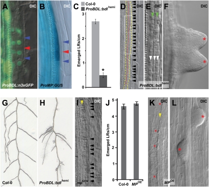
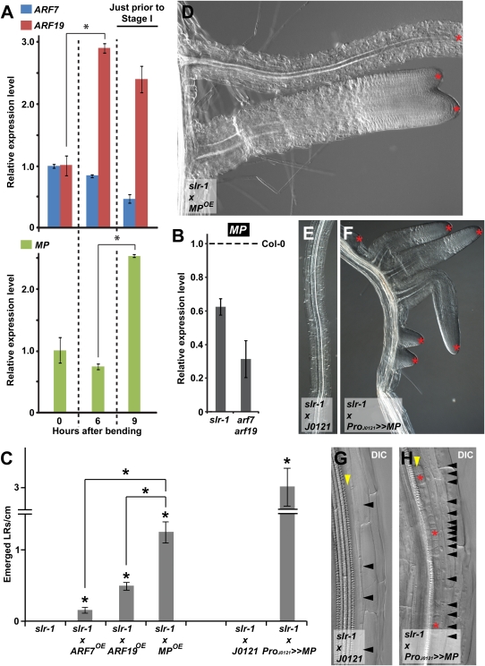
Like animals, the mature plant body develops via successive sets of instructions that determine cell fate, patterning, and organogenesis. In the coordination of various developmental programs, several plant hormones play decisive roles, among which auxin is the best-documented hormonal signal. Despite the broad range of processes influenced by auxin, how such a single signaling molecule can be translated into a multitude of distinct responses remains unclear. In Arabidopsis thaliana, lateral root development is a classic example of a developmental process that is controlled by auxin at multiple stages. Therefore, we used lateral root formation as a model system to gain insight into the multifunctionality of auxin. We were able to demonstrate the complementary and sequential action of two discrete auxin response modules, the previously described Solitary Root/indole-3-Acetic Acid (IAA)14-Auxin Response Factor (ARF)7-ARF19-dependent lateral root initiation module and the successive Bodenlos/IAA12-Monopteros/ARF5-dependent module, both of which are required for proper organogenesis. The genetic framework in which two successive auxin response modules control early steps of a developmental process adds an extra dimension to the complexity of auxin's action.


Conflict of interest statement

The authors declare no conflict of interest.

Figures

Fig. 1.
Fig. 1.
Auxin response module after cell cycle activation. (A) WT (Col-0) lateral root initiation. Arrowheads indicate adjacent (red) and short cell boundaries (blue). (B and C) Pericycle cell boundaries (black arrowheads) in a Col-0 (B) and E2Fa/DPaOE (C) root regions. The yellow arrowhead indicates single pericycle layer. DIC, differential interference contrast (Nomarski) imaging. (Scale bars: 50 μM.) (D) Number of lateral roots/cm in WT (Col-0 and Ler), CYCD3,1OE, and E2Fa/DPaOE (mean ± SEM) at different NAA concentrations. Seedlings 5 days after germination were transferred to NAA for 5 days. *Statistically significant differences for values compared with WT as determined by Student's t test (P < 0.05).
Fig. 2.
Fig. 2.
Auxin response module after SLR-dependent cell cycle activation. Absence of lateral roots (A–C), lack of expression of the marker ProACR4:H2B:YFP (G–I), and lack of increased expression of PLT3 (K) in slr-1 (A, G, and K), slr-1 grown on NAA (B, H, and K), and slr-1 with increased cell cycle activity (slr-1xCYCD3;1OE) (C, I, and K), but presence of stretches of short cells in slr-1xCYCD3;1OE (D). Induction of lateral roots (E and F) and/or turning on of ProACR4:H2B:YFP (J) and PLT3 (K) in slr-1xCYCD3;1OE (E, J, and K) and slr-1xE2Fa/DPaOE (F) by the combination of NAA and increased cell cycle activity. Insets (G, and I) show the presence of ProACR4:H2B:YFP in the root tip. Seedlings 5 days after germination were transferred to 10 μM NAA for 96 h (B, E, and F) and 30 h (H and J). Arrowheads indicate the pericycle (yellow) (D and J), which consists of one layer (D) or of one layer starting to undergo periclinal divisions (J) and pericycle cell boundaries (black) (D). DIC, differential interference contrast (Nomarski) imaging. (K) Quantitative RT-PCR expression analysis of PLT3. Seedlings 5 days after germination were treated with 10 μM NAA for 24 h.
Fig. 3.
Fig. 3.
Role of BDL and MP in lateral root development. (A and B) Expression of ProBDL:n3xGFP (A) and ProMP:GUS (B) in the lateral root initiation site. Arrowheads indicate adjacent (red) and short cell boundaries (blue). (C–L) bdl gain of function [hemizygous ProBDL:bdl:GUS (ProBDL:bdlhemi)] (C–F and H), mp partial loss of function (mpS319) (I), and MP overexpression (MPOE) (J–L) leading to defects in pericycle cell division, lateral root initiation, and positioning compared with WT (Col-0) (C, G, and J). (C and J) Emerged lateral root density at 10 (J) and 11 (C) days after germination. (G and H) Lateral root positioning in Col-0 WT (G) and ProBDL:bdlhemi (H) at 15 days after germination. The red dotted line indicates separation between two pericycle cell layers, and the yellow dashed line indicates outer boundaries of the two- or three-layer pericycle (D). Arrowheads indicate two- (green) and three-layer pericycle (white) (E), small pericycle cell boundaries (black) (D and I), and pericycle (yellow), which is undergoing periclinal division (I) or consists of one layer (K). Red asterisks indicate lateral root primordia (F and L) and sites that strongly resemble a lateral root initiation site (K). Data are mean ± SEM. *Statistically significant differences for values compared with WT as determined by Student's t test (P < 0.01).
Fig. 4.
Fig. 4.
BDL-MP–dependent auxin response module after SLR/IAA14-ARF7-ARF19 action. (A and B) Quantitative RT-PCR expression analysis of ARF7, ARF19, and MP during synchronized lateral root initiation (A) and of MP in slr-1 (5 days after germination) and arf7arf19 roots (10 days after germination) compared with WT expression level (dashed line) (B), indicating two successive modules. Just prior to stage I in (A) indicates at which time point CYCB1;1 expression is induced, which coincides with the onset of lateral root initiation. (CH) Induction of lateral roots in slr-1 by expression in whole roots MPOE (C and D) and pericycle-specific expression (ProJ0121>>MP) of MP (C and E–H). (C) Emerged lateral root density at 12 days (slr-1, slr-1xMPOE, slr-1xARF7OE, and slr-1xARF19OE) and 15 days (slr-1xJ0121 and slr-1xProJ0121>>MP) after germination. Data are mean ± SEM. *Statistically significant differences for values compared with control or as indicated with a black line as determined by Student’s t test (P < 0.01). (D–H) Red asterisks indicate emerged lateral roots (D and F) and sites that strongly resemble a lateral root initiation site (H) in slr-1xMPOE (D) and in slr-1xProJ0121>>MP (F and H) compared with the control slr-1xJ0121 (E and G). Arrowheads indicate pericycle cell boundaries (black) (G and H) and a pericycle consisting of a single layer (yellow) (G and H). DIC, differential interference contrast (Nomarski) imaging.

Similar articles

Cited by

References

    1. Vanneste S, Friml J. Auxin: A trigger for change in plant development. Cell. 2009;136:1005–1016. - PubMed
    1. De Smet I, et al. Receptor-like kinase ACR4 restricts formative cell divisions in the Arabidopsis root. Science. 2008;322:594–597. - PubMed
    1. Péret B, et al. Arabidopsis lateral root development: An emerging story. Trends Plant Sci. 2009;14:399–408. - PubMed
    1. Fukaki H, Tasaka M. Hormone interactions during lateral root formation. Plant Mol Biol. 2009;69:437–449. - PubMed
    1. Lau S, Jürgens G, De Smet I. The evolving complexity of the auxin pathway. Plant Cell. 2008;20:1738–1746. - PMC - PubMed

Publication types

MeSH terms

LinkOut - more resources