. 2010 Feb 9;107(6):2574-9.
doi: 10.1073/pnas.0915018107. Epub 2010 Jan 25.

Differential release of chromatin-bound IL-1alpha discriminates between necrotic and apoptotic cell death by the ability to induce sterile inflammation

Affiliations

Differential release of chromatin-bound IL-1alpha discriminates between necrotic and apoptotic cell death by the ability to induce sterile inflammation

Idan Cohen et al. Proc Natl Acad Sci U S A. .

Abstract

IL-1alpha, like IL-1beta, possesses multiple inflammatory and immune properties. However, unlike IL-1beta, the cytokine is present intracellularly in healthy tissues and is not actively secreted. Rather, IL-1alpha translocates to the nucleus and participates in transcription. Here we show that intracellular IL-1alpha is a chromatin-associated cytokine and highly dynamic in the nucleus of living cells. During apoptosis, IL-1alpha concentrates in dense nuclear foci, which markedly reduces its mobile nature. In apoptotic cells, IL-1alpha is retained within the chromatin fraction and is not released along with the cytoplasmic contents. To simulate the in vivo inflammatory response to cells undergoing different mechanisms of death, lysates of cells were embedded in Matrigel plugs and implanted into mice. Lysates from cells undergoing necrosis recruited cells of the myeloid lineage into the Matrigel, whereas lysates of necrotic cells lacking IL-1alpha failed to recruit an infiltrate. In contrast, lysates of cells undergoing apoptotic death were inactive. Cells infiltrating the Matrigel were due to low concentrations (20-50 pg) of the IL-1alpha precursor containing the receptor interacting C-terminal, whereas the N-terminal propiece containing the nuclear localization site failed to do so. When normal keratinocytes were subjected to hypoxia, the constitutive IL-1alpha precursor was released into the supernatant. Thus, after an ischemic event, the IL-1alpha precursor is released by hypoxic cells and incites an inflammatory response by recruiting myeloid cells into the area. Tissues surrounding the necrotic site also sustain damage from the myeloid cells. Nuclear trafficking and differential release during necrosis vs. apoptosis demonstrate that inflammation by IL-1alpha is tightly controlled.


Conflict of interest statement

The authors declare no conflict of interest.

Figures
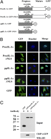
Fig. 1.
Fig. 1.
IL-1α is a chromatin-associated cytokine. (A) Schematic representation of IL-1α GFP-conjugated proteins. The N-terminal domain containing the NLS in position 82–89 is denoted by a dark gray box. The C-terminal mature component (known as the IL-1R interacting domain) is represented by a clear box. (B) B16 melanoma cells were transfected with precIL-1α, ppIL-1α, and IL-1α mutants containing inactive NLS or GFP. Twenty-four hours later, nuclear localization and chromatin alignment were analyzed by confocal microscopy. (C) ChIP assays were performed using sheared chromatin from lysates of precIL-1α or GFP (as a control) transfected cells and were immunoprecipitated with anti-GFP antibody. An additional control assay was performed without the presence of antibody (beads only). The immunoprecipitates were separated over a 4–20% gradient SDS/PAGE gel and transferred to a nitrocellulose membrane. To verify chromatin presence, histone H3 antibody served as a molecular marker in Western blot analysis.
Fig. 2.
Fig. 2.
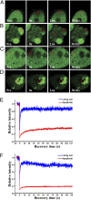
Dynamics of IL-1α nuclear proteins in living and apoptotic cells. FRAP experiments in living and apoptotic cells expressing ppIL-1α-GFP and precIL-1α-GFP. (A) Bleached region of ppIL-1α-GFP–transfected B16 cell. (B) Bleached region of UV-irradiated ppIL-1α-GFP–transfected B16 cell. (C) Bleached region of precIL-1α-GFP–transfected B16 cell. (D) Bleached region of UV-irradiated precIL-1α-GFP–transfected B16 cell. Images were taken at the indicated times before (Pre), during (0s), and after the end of the bleach pulse (2.6s and 30.01s). The area of the bleach spot is indicated with a red circle. (E) Kinetics of nucleoplasmic FRAP in living or UV-irradiated apoptotic B16 melanoma cells transfected with ppIL-1α-GFP. (F) Kinetics of nucleoplasmic FRAP in living or UV-irradiated apoptotic B16 melanoma cells transfected with precIL-1α-GFP. Fluorescence intensity in the bleached region was measured for at least 110 s and expressed as the relative recovery. Data values in FRAP kinetics represent one of six independent experiments.
Fig. 3.
Fig. 3.
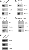
Differential release of IL-1α by necrotic and apoptotic cells. The release of IL-1α and β-actin from precIL-1α-GFP–transfected B16 melanoma cells was determined by Western blot analysis, using the denoted antibodies. Histones were visualized using Ponceau S staining. (A) For the secretion of IL-1α and β-actin from intact live cells, the cells and the culture medium were analyzed by Western blot. Cell lysates from necrotic cells (B) or apoptotic cells (C) were obtained by three cycles of freeze–thawing and further fractionated by centrifugation. The soluble (S) and nonsoluble (P) fractions were analyzed as described above. (D) Apoptotic pellets were further treated with 1% Nonidet P-40 for 1 h, and both the soluble and nonsoluble fractions were analyzed. (E) Soluble fractions from necrotic and apoptotic (described in B and C) cells were analyzed for the presence of β-actin and the nuclear proteins Suz12 and Ezh2, using the appropriate antibodies.
Fig. 4.
Fig. 4.
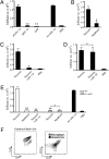
IL-1α release from dying cells distinguishes between necrosis and apoptosis by triggering a proinflammatory response. (A) Number of infiltrating cells into Matrigel plugs 20 h after injection with supernatants of necrotic ppIL-1α, precIL-1α, or GFP-transfected B16 melanoma cells. Recombinant mIL-1α and PBS were used as positive and negative controls, respectively. *P < 0.05, **P < 0.01 vs. the precIL-1α group. (B) Infiltrating cell numbers in Matrigel plugs 20 h after injection with supernatants of necrotic and apoptotic precIL-1α–transfected B16 melanoma cells. *P < 0.05, **P < 0.01 vs. the necrosis group. (C and D) Infiltrating cell numbers in Matrigel plugs 20 h after injection with supernatants of necrotic precIL-1α–transfected B16 melanoma cells with or without neutralizing anti-IL-1α antibodies (C) or anti-IL-1β antibodies (D). *P < 0.05, **P < 0.01 vs. the necrosis group. (E) Necrotic and apoptotic WT and IL-1α−/− fibroblasts were injected with or without neutralizing anti-IL-1α antibodies together with Matrigel. Graph represents infiltrating cell numbers after 20 h. *P < 0.05, **P < 0.01 vs. the necrosis group. (F) FACS analysis of infiltrating cells into Matrigel plugs. Macrophages were defined as Ly-6Chigh/CD115+/F4-80+ cells; neutrophils were defined as Ly-6G high/Ly-6Cdull/CD115neg cells. All experiments described in this figure were repeated three times, and each experimental group consisted of three mice. Data are from a single experiment and are presented as average ± SD.
Fig. 5.
Fig. 5.
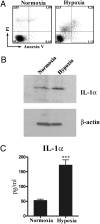
The precursor of IL-1α is released from necrotic cells after hypoxia. (A) FACS analysis of annexin-V/PI–stained BD7 cells after 24 h culture in normoxic (Left) or hypoxic conditions (Right). (B) Lysates of the BD7 cells were analyzed by Western blot for IL-1α (Top) 24 h after culture in normoxic or hypoxic conditions, showing the 31-kDa precursor form. β-actin was used as loading control (Bottom). (C) Concentration of IL-1α in culture media of BD7 keratinocytes after 24 h culture in normoxic or hypoxic conditions, as measured by ELISA. Data represents the average ± SD (n = 3). ***P = 0.0005.

Similar articles

Cited by

References

    1. Apte RN, Voronov E. Is interleukin-1 a good or bad ‘guy’ in tumor immunobiology and immunotherapy? Immunol Rev. 2008;222:222–241. - PubMed
    1. Dinarello CA. Immunological and inflammatory functions of the interleukin-1 family. Annu Rev Immunol. 2009;27:519–550. - PubMed
    1. Mosley B, et al. The interleukin-1 receptor binds the human interleukin-1 alpha precursor but not the interleukin-1 beta precursor. J Biol Chem. 1987;262:2941–2944. - PubMed
    1. Buryskova M, Pospisek M, Grothey A, Simmet T, Burysek L. Intracellular interleukin-1alpha functionally interacts with histone acetyltransferase complexes. J Biol Chem. 2004;279:4017–4026. - PubMed
    1. Werman A, et al. The precursor form of IL-1alpha is an intracrine proinflammatory activator of transcription. Proc Natl Acad Sci USA. 2004;101:2434–2439. - PMC - PubMed

Publication types

MeSH terms