Clinical Trial
. 2010 Feb 9;107(6):2681-6.
doi: 10.1073/pnas.0913008107. Epub 2010 Jan 25.

Breakdown in cortical effective connectivity during midazolam-induced loss of consciousness

Affiliations
Clinical Trial

Breakdown in cortical effective connectivity during midazolam-induced loss of consciousness

Fabio Ferrarelli et al. Proc Natl Acad Sci U S A. .

Abstract

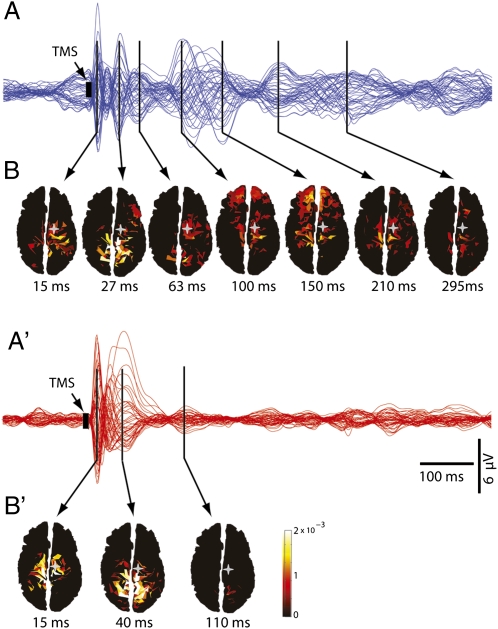
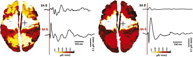
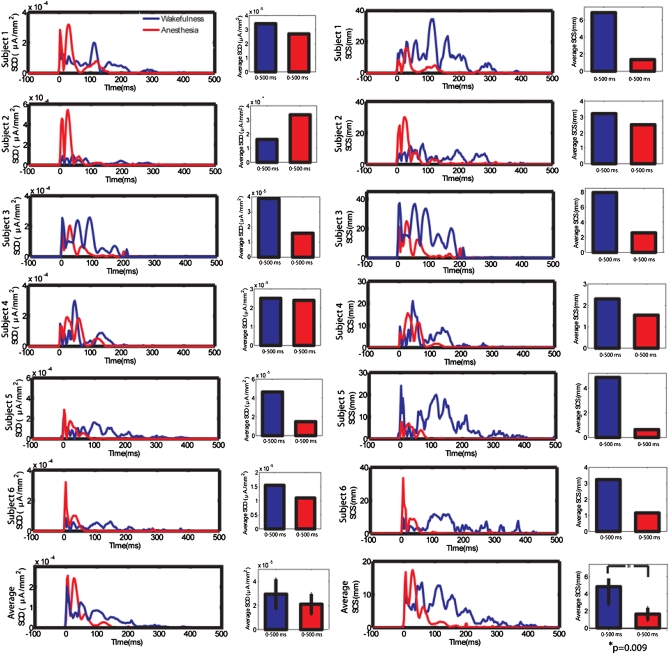
By employing transcranial magnetic stimulation (TMS) in combination with high-density electroencephalography (EEG), we recently reported that cortical effective connectivity is disrupted during early non-rapid eye movement (NREM) sleep. This is a time when subjects, if awakened, may report little or no conscious content. We hypothesized that a similar breakdown of cortical effective connectivity may underlie loss of consciousness (LOC) induced by pharmacologic agents. Here, we tested this hypothesis by comparing EEG responses to TMS during wakefulness and LOC induced by the benzodiazepine midazolam. Unlike spontaneous sleep states, a subject's level of vigilance can be monitored repeatedly during pharmacological LOC. We found that, unlike during wakefulness, wherein TMS triggered responses in multiple cortical areas lasting for >300 ms, during midazolam-induced LOC, TMS-evoked activity was local and of shorter duration. Furthermore, a measure of the propagation of evoked cortical currents (significant current scattering, SCS) could reliably discriminate between consciousness and LOC. These results resemble those observed in early NREM sleep and suggest that a breakdown of cortical effective connectivity may be a common feature of conditions characterized by LOC. Moreover, these results suggest that it might be possible to use TMS-EEG to assess consciousness during anesthesia and in pathological conditions, such as coma, vegetative state, and minimally conscious state.


Conflict of interest statement

The authors declare no conflict of interest.

Figures

Fig. 1.
Fig. 1.
Spatiotemporal dynamics of TMS-evoked activity change markedly during LOC. (A and A′) Averaged TMS-evoked potentials at all electrodes, superimposed in butterfly plots (blue traces for waking, red traces for anesthesia). (B and B′) Cortical currents calculated on individual cortical meshes are shown from minimal (dark red) to maximal (white) values. During wakefulness, TMS of premotor cortex determined low-amplitude, complex scalp waves corresponding to cortical currents that lasted >300 ms and shifted among distant cortical areas. Conversely, during anesthesia, TMS gave rise to high-amplitude, short-lasting scalp voltages reflecting cortical currents that remained local, and faded within 150 ms. Gray stars, TMS target (premotor cortex); black arrows, local maxima in periods of significant TMS-evoked activation.
Fig. 2.
Fig. 2.
TMS during anesthesia evokes a large positive–negative wave in the stimulation site but little activation in distant areas. Cortical currents evoked by TMS of premotor cortex, cumulated in a 0–500 ms post-TMS interval and displayed on the corresponding Broadmann areas (BA) in wakefulness (Left) and anesthesia (Right). To the right of each topographic plot are time courses of currents recorded from the stimulated area, premotor cortex (BA 6), and from a more anterior cortical area (BA 8). During anesthesia, TMS-evoked SCD in BA6 was similar to the SCD recorded in wakefulness, as reflective of an initial stronger but shorter-lived response during anesthesia compared to wakefulness. Conversely, SCD from BA 8, which is anatomically connected to BA 6, were markedly reduced in anesthesia compared to wakefulness, suggesting a marked decrease in cortical effective connectivity.
Fig. 3.
Fig. 3.
A synthetic index of cortical connectivity (SCS), but not reactivity (SCD), captures cortical changes during LOC. SCD and SCS were computed for each subject in wakefulness (blue line) and anesthesia (red line) following TMS of the premotor cortex. (Left) Time course of SCD for individual and average data, and mean SCD over the entire post-TMS time interval (0–500 ms). In each subject, SCD values were initially higher (first 50 ms after TMS) during anesthesia but tended to dissipate shortly thereafter, consistent with a TMS-evoked larger initial response during anesthesia that was, however, short-lived (Fig. 1 A′ and B′). Mean SCD over the entire post-TMS period were not significantly different between wakefulness and anesthesia. (Right) Time course of SCS for individual and average data, and mean SCS over the entire post-TMS interval. In each subject, during wakefulness, SCS was present for >200 ms, whereas during anesthesia it faded after 100 ms. Notably, mean SCS values in the 0–500 ms post-TMS interval were significantly higher in wakefulness relative to anesthesia (P = 0.009, Mann–Whitney).

References

    1. Tononi G. An information integration theory of consciousness. BMC Neurosci. 2004;5:42–62. - PMC - PubMed
    1. Hobson JA, Pace-Schott EF. The cognitive neuroscience of sleep: neuronal systems, consciousness and learning. Nat Rev Neurosci. 2002;3:679–693. - PubMed
    1. Steriade M, Timofeev I, Grenier F. Natural waking and sleep states: a view from inside neocortical neurons. J Neurophysiol. 2001;85:1969–1985. - PubMed
    1. Ilmoniemi RJ, et al. Neuronal responses to magnetic stimulation reveal cortical reactivity and connectivity. Neuroreport. 1997;8:3537–3540. - PubMed
    1. Massimini M, et al. Breakdown of cortical effective connectivity during sleep. Science. 2005;309:2228–2232. - PubMed

Publication types