. 2010 Feb 9;107(6):2658-63.
doi: 10.1073/pnas.0914957107. Epub 2010 Jan 25.

A role for FKBP52 in Tau protein function

Affiliations

A role for FKBP52 in Tau protein function

Béatrice Chambraud et al. Proc Natl Acad Sci U S A. .

Abstract

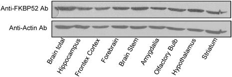
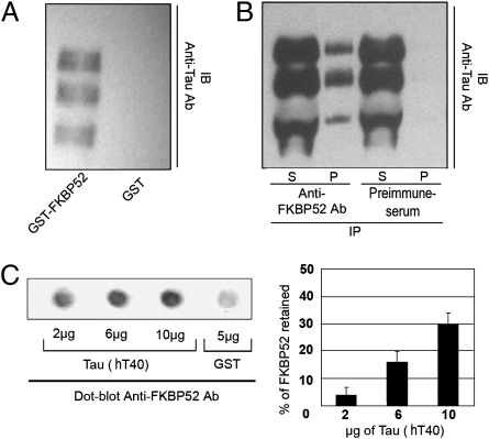
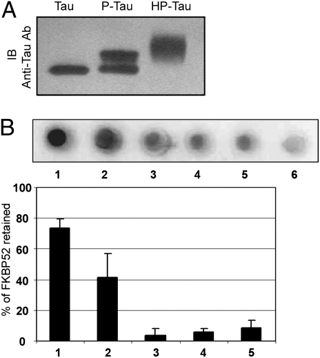
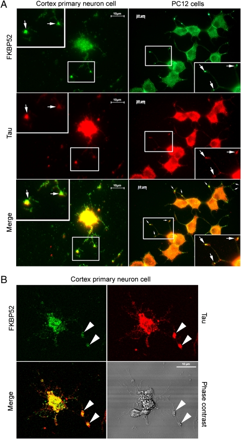
Tau is a microtubule-associated protein, which is widely expressed in the central nervous system, predominantly in neurons, where it regulates microtubule dynamics, axonal transport, and neurite outgrowth. The aberrant assembly of Tau is the hallmark of several human neurodegenerative diseases, collectively known as tauopathies. They include Alzheimer's disease, Pick's disease, progressive supranuclear palsy, and frontotemporal dementia and parkinsonism linked to chromosome 17. Several abnormalities in Tau, such as hyperphosphorylation and aggregation, alter its function and are central to the pathogenic process. Here, we describe biochemical and functional interactions between FKBP52 and Tau. FKBP52 is a member of the FKBP (FK506-binding protein) family that comprises intracellular protein effectors of immunosuppressive drugs (such as FK506 and rapamycin). We found that FKBP52, which is abundant in brain, binds directly and specifically to Tau, especially in its hyperphosphorylated form. The relevance of this observation was confirmed by the colocalization of both proteins in the distal part of the axons of cortical neurons and by the antagonistic effect of FKBP52 on the ability of Tau to promote microtubule assembly. Overexpression of FKBP52 in differentiated PC12 cells prevented the accumulation of Tau and resulted in reduced neurite length. Taken together, these findings indicate a role for FKBP52 in Tau function and may help to decipher and modulate the events involved in Tau-induced neurodegeneration.


Conflict of interest statement

The authors declare no conflict of interest.

Figures

Fig. 1.
Fig. 1.
FKBP52 in the brain. Twenty micrograms of cytosol proteins from different adult rat brain regions were analyzed by Western blotting using anti-FKBP52 antibody 761. Actin served as the loading control.
Fig. 2.
Fig. 2.
Association between FKBP52 and Tau proteins. (A) GST pull-down assay: immunoblot (IB) for Tau showing the binding of soluble microtubule extract proteins incubated with GST-tagged FKBP52 or GST alone as control. (B) Coimmunoprecipitation assay: a soluble microtubule extract was subjected to immunoprecipitation (IP) with immunopurified anti-FKBP52 antibody or preimmune serum used as control. The supernatants (S) and the precipitates (P) were immunoblotted with anti-Tau antibody (clone DC25). (C) The ability of Tau proteins to bind FKBP52 directly was monitored by dot blot assay. Different amounts of recombinant Tau (hT40) were spotted onto nitrocellulose membranes and then assayed for bound FKBP52 (0.5 μg) using anti-FKBP52 antibody. Five micrograms of GST spotted onto nitrocellulose membrane was used as the control. Quantitation: 100% corresponds to 0.5 μg of FKBP52 loading before milk saturation, and 0% corresponds to 0.5 μg of FKBP52 loading after milk saturation. The background is defined as the signal when GST instead of hT40 was loaded. The level of FKBP52 captured by hT40 was calculated after substraction of background.
Fig. 3.
Fig. 3.
Relevance of Tau phosphorylation for its interaction with FKBP52. (A) Recombinant Tau (hT40), P-Tau, and HP-Tau were analyzed by SDS–Page. Phosphorylation and hyperphosphorylation of hT40 resulted in a marked reduction in the gel mobility of recombinant Tau as shown on an immunoblot (IB) with anti-Tau antibody (clone DC25). (B) Dot blot assay with 2.2 μg of HP-Tau (1), P-Tau (2), and pure recombinant hT40 (3) to which had been added, just before spotting, the same amount of cytosol as used to generate P-Tau (4) or HP-Tau (5). Lane 6 shows the GST (5 μg) load.
Fig. 4.
Fig. 4.
Colocalization of FKBP52 and Tau in primary cortical neurons and PC12 cells. (A) Immunofluorescence staining of primary cortical neurons and PC12 cells treated with 50 nM NGF for 5 days. Double staining for Tau and FKBP52 was performed after cytosol extraction to reveal cytoskeletal association. Arrows indicate preferential colocalization of both proteins in the distal part of the nerve cell axon and at the extremity of PC12 cell neurites. (B) Confocal images of primary cortical neurons. Double staining was performed as in A. Analysis of 0.5-μm slices confirms the preferential colocalization in the distal part of the axon (arrowheads).
Fig. 5.
Fig. 5.
Effect of FKBP52 on tubulin polymerization induced by recombinant Tau isoforms. Tubulin polymerization was performed by switching the samples from 4 °C to 37 °C, and the change in turbidity was monitored at 345 nm for 15 min. Tubulin (1 mg/mL) purified from rat brain was incubated in the absence (formula image
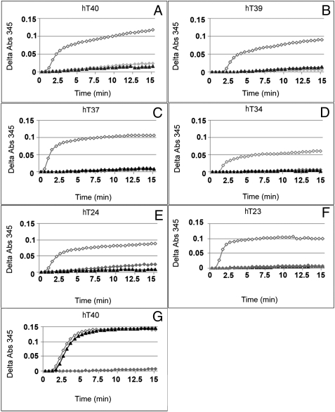
) or presence of 1.7 μM (23 μg for HT40) different human Tau isoforms (as indicated in panels A‒F) without FKBP52 (◇) or with 3.5 μM (55 μg) FKBP52 (▲). Tau isoforms differ from each other by the number of repeats in the microtubule-binding domain and insertions in the N terminus. The labeling of Tau isoforms uses the published nomenclature (21). (G) This control experiment was carried out as in A, except that 3.5 μM GST (▲) was used instead of FKBP52.
Fig. 6.
Fig. 6.
Effect of FKBP52 overexpression on Tau accumulation and neurite outgrowth. (A) The FKBP52 level was determined by Western blot using anti-FKBP52 antibody 761, in H7C2 cells treated or not with Dox. The use of rabbit FKBP52 as the exogenous protein explains the small difference in gel mobility with the endogenous rat protein. (B) PC12 cells treated or not with NGF (50 nM) in the absence or presence of 1 μg/mL Dox (doxycycline). Ten micrograms of total protein extracts were analyzed for FKBP52 levels as in A. (C) H7C2 and PC12 cells were treated or not with NGF for 5 days in the presence or absence of Dox for 1 week; 50 μg of extracts was subjected to SDS–PAGE and immunoblotted with anti-Tau (antibody clone DC25). Actin was used as the loading control. (D) Representative H7C2 cells in the presence of NGF (50 nM) with or without Dox. Neurite length was quantified from random photographs (Materials and Methods). Similar results were obtained in three separate experiments. **P < 0.01 (Student–Newman–Keuls test, ANOVA).

Similar articles

Cited by

References

    1. Schreiber SL. Chemistry and biology of the immunophilins and their immunosuppressive ligands. Science. 1991;251:283–287. - PubMed
    1. Steiner JP, et al. High brain densities of the immunophilin FKBP colocalized with calcineurin. Nature. 1992;358:584–587. - PubMed
    1. Snyder SH, Sabatini DM. Immunophilins and the nervous system. Nat Med. 1995;1:32–37. - PubMed
    1. Gold BG. FK506 and the role of immunophilins in nerve regeneration. Mol Neurobiol. 1997;15:285–306. - PubMed
    1. Shim S, et al. Peptidyl-prolyl isomerase FKBP52 controls chemotropic guidance of neuronal growth cones via regulation of TRPC1 channel opening. Neuron. 2009;64:471–483. - PMC - PubMed

Publication types

MeSH terms