. 2010 Apr;176(4):2029-38.
doi: 10.2353/ajpath.2010.090755. Epub 2010 Feb 4.

Novel EphB4 monoclonal antibodies modulate angiogenesis and inhibit tumor growth

Affiliations

Novel EphB4 monoclonal antibodies modulate angiogenesis and inhibit tumor growth

Valery Krasnoperov et al. Am J Pathol. 2010 Apr.

Abstract

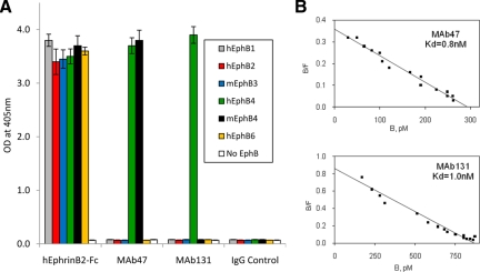
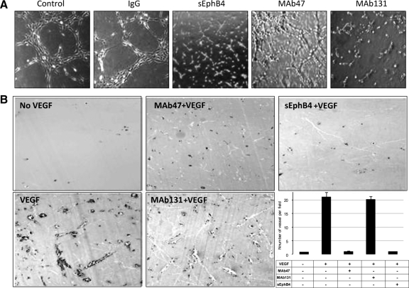
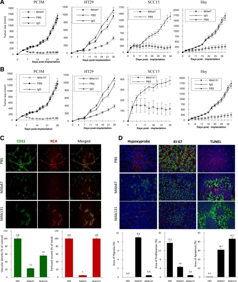
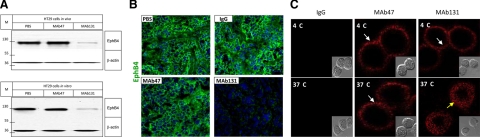
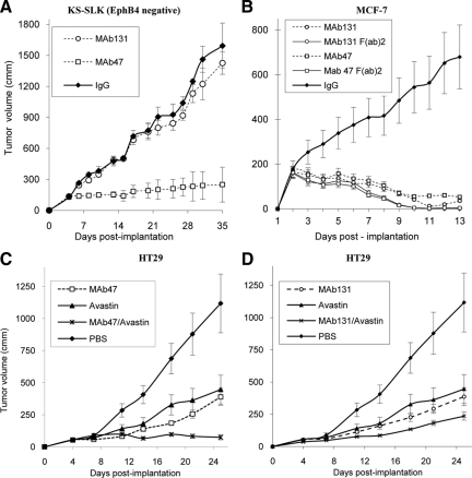
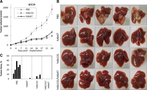
EphB4 receptor tyrosine kinase and its cognate ligand EphrinB2 regulate induction and maturation of newly forming vessels. Inhibition of their interaction arrests angiogenesis, vessel maturation, and pericyte recruitment. In addition, EphB4 is expressed in the vast majority of epithelial cancers and provides a survival advantage to most. Here, we describe two anti-EphB4 monoclonal antibodies that inhibit tumor angiogenesis and tumor growth by two distinct pathways. MAb131 binds to fibronectin-like domain 1 and induces degradation of human EphB4, but not murine EphB4. MAb131 inhibits human endothelial tube formation in vitro and growth of human tumors expressing EphB4 in vivo. In contrast, MAb47 targets fibronectin-like domain 2 of both human and murine EphB4 and does not alter EphB4 receptor levels, but inhibits angiogenesis and growth of both EphB4-positive and EphB4-negative tumors in a mouse s.c. xenograft model. Combination of MAb47 and bevacizumab enhances the antitumor activity and induces tumor regression. Indeed, humanized antibodies hAb47 and hAb131 showed similar affinity for EphB4 and retained efficacy in the inhibition of primary tumor development and experimental metastasis.


Figures

Figure 1
Figure 1
Binding of monoclonal antibodies to EphB4. A: MAbs were immobilized on Protein A-Agarose beads and incubated with indicated recombinant EphB ECDs of human (h) or mouse (m) origin fused to AP. Negative controls included beads with AP alone (No EphB) and beads with unrelated IgG (IgG Control). As a positive control hEphrinB2-ECD fused to Fc was used in place of antibodies. B: To measure antibody affinity, biotinylated MAb47 (top) or MAb131 (bottom) were immobilized on Streptavidin-Agarose beads. Different concentrations (50 to 5000 pmol/L) of hEphB4-ECD-AP were applied to obtain saturation plot, nonspecific binding (no MAb added) values were subtracted and converted into coordinates of Scatchard plot.
Figure 2
Figure 2
Effect of antibodies on human umbilical vein endothelial tube formation assay and Matrigel plug assays: A: Human umbilical vein endothelial cells (20,000 per well) were premixed with test compounds at the final concentration of 10 μg/ml and plated on matrigel coated wells. Negative controls included PBS or unrelated mouse IgG. Positive control consisted of soluble EphB4 (sEphB4) applied at a concentration of 5 μg/ml (×20). Pictures were taken using a Bioquant Image Analysis system. B: Matrigel impregnated with test compounds (0.5 ml) was implanted in mouse abdominal wall. Plugs were harvested on day 7 and analyzed for vessel density (×10). Quantification of vessel density was done using image analysis system. Data are presented as mean ± SE.
Figure 3
Figure 3
Human tumor xenograft studies in vivo. A and B: Mice were implanted subcutaneously with EphB4-positive cell lines; mice with established tumors were randomized to different treatment groups. Tumor volumes over time after treatment with MAb47 (A) or MAb131 (B) are shown. Controls include PBS or isotype specific IgG. C: Analysis of HT29 tumor tissues harvested after therapy. Representative data for vessel density (CD31 staining in green), vessel perfusion (RCA in red), and merged images (×20) are shown. Quantification of vessel density and perfusion was done using Bioquant image analysis. Data are presented as mean ± SE. D: Tissue analysis of HT29 tumors was performed for areas of hypoxia (green), proliferative index by Ki-67 staining (green), and apoptosis by TUNEL assay (green). Perfused vessels were identified by RCA localization (red). Quantification of areas of hypoxia, proliferation, and apoptosis was done using Bioquant image analysis (×20). Data are presented as mean ± SE.
Figure 4
Figure 4
EphB4 degradation and endocytosis after antibody treatment. A: Western blots for level of EphB4 in HT29 tumor tissue harvested at the end of in vivo experiments (top) or in HT29 cells treated in vitro at a dose of 10 μg/ml for 12 hours (bottom) are shown. “M” represents molecular mass marker in kDa. B: Immunofluorescence assay for EphB4 localization (green) and nuclear staining with DAPI (blue) was performed on HT29 tumor xenografts at the end of the antibody therapy. C: HT29 cells were exposed to Cy5 labeled IgG, MAb47, or MAb131 at 4°C or 37°C for 30 minutes. EphB4 was localized (red) using confocal microscope. MAb131 at 37°C induces endocytosis (yellow arrow), while MAb47 does not (white arrow). No endocytosis is observed at 4°C (white arrows) with eithor antibody. Cells imaging using differential interference contrast are shown in the insets.
Figure 5
Figure 5
A: MAb therapy of EphB4-negative tumor xenografts. EphB4-negative KS-SLK cell line was implanted and tumor xenografts were treated with MAb47, MAb131, or IgG as in Figure 3, A and B. Tumor volumes were measured over time. B: Activity of full-length EphB4 antibodies (twice a week i.p.) or F(ab)2 fragments (three times a week i.p.) were tested in mice bearing HT29 tumors as in Figure 3, A and B. C and D: Combination studies of EphB4 antibodies and VEGF antibody. HT29 tumor-bearing mice were assigned to treatment groups of MAb47, MAb131, and VEGF antibody (Avastin) each one alone, or combination of Avastin plus MAb47 or Avastin plus MAb131. In combination studies, the dose of each antibody was reduced by 50% (5 mg/kg). Tumor volumes were measured over time.
Figure 6
Figure 6
Activity of Humanized MAbs in vivo. A: HT29 tumor xenografts in nu/nu mice were treated with either hAb47 or hAb131 i.p. three times a week, at the dose of 10 mg/kg. Tumor volumes were measured over time. B: Activity of humanized EphB4 antibodies in liver metastasis after intrasplenic injection of HT29 colon ca cell line. hAb47 and hAb131 treatment was administered i.p. three times a week. Images of livers harvested at the end of the study from each treatment group are shown. C: Area of the liver occupied by the tumor is provided.

References

    1. Griffioen AW, Molema G. Angiogenesis: potentials for pharmacologic intervention in the treatment of cancer, cardiovascular diseases, and chronic inflammation. Pharmacol Rev. 2000;52:237–268. - PubMed
    1. Yancopoulos GD, Davis S, Gale NW, Rudge JS, Wiegand SJ, Holash J. Vascular-specific growth factors and blood vessel formation. Nature. 2000;407:242–248. - PubMed
    1. Kerbel RS. Tumor angiogenesis. N Engl J Med. 2008;358:2039–2049. - PMC - PubMed
    1. Brantley-Sieders DM, Chen J. Eph receptor tyrosine kinases in angiogenesis: from development to disease. Angiogenesis. 2004;7:17–28. - PubMed
    1. Wang HU, Chen ZF, Anderson DJ. Molecular distinction and angiogenic interaction between embryonic arteries and veins revealed by ephrin-B2 and its receptor Eph-B4. Cell. 1998;93:741–753. - PubMed

Publication types