. 2010 Feb 2;107(5):1971-6.
doi: 10.1073/pnas.0908569107. Epub 2010 Jan 19.

Crystal structure of the DNA-recognition component of the bacterial virus Sf6 genome-packaging machine

Affiliations

Crystal structure of the DNA-recognition component of the bacterial virus Sf6 genome-packaging machine

Haiyan Zhao et al. Proc Natl Acad Sci U S A. .

Abstract

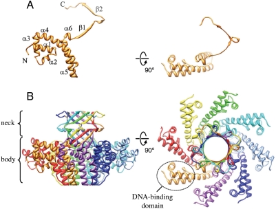
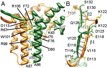
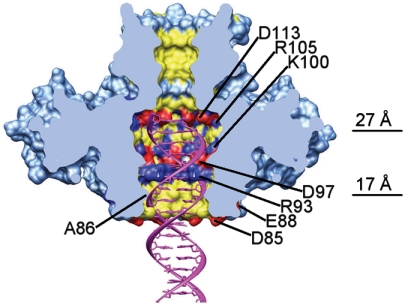
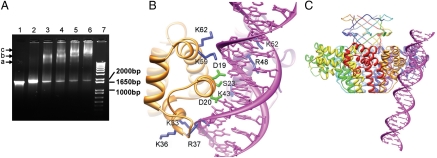
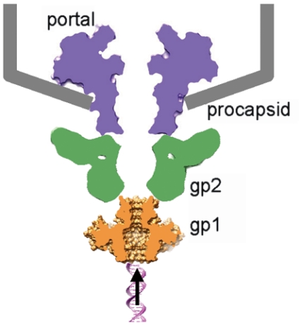
In herpesviruses and many bacterial viruses, genome-packaging is a precisely mediated process fulfilled by a virally encoded molecular machine called terminase that consists of two protein components: A DNA-recognition component that defines the specificity for packaged DNA, and a catalytic component that provides energy for the packaging reaction by hydrolyzing ATP. The terminase docks onto the portal protein complex embedded in a single vertex of a preformed viral protein shell called procapsid, and pumps the viral DNA into the procapsid through a conduit formed by the portal. Here we report the 1.65 A resolution structure of the DNA-recognition component gp1 of the Shigella bacteriophage Sf6 genome-packaging machine. The structure reveals a ring-like octamer formed by interweaved protein monomers with a highly extended fold, embracing a tunnel through which DNA may be translocated. The N-terminal DNA-binding domains form the peripheral appendages surrounding the octamer. The central domain contributes to oligomerization through interactions of bundled helices. The C-terminal domain forms a barrel with parallel beta-strands. The structure reveals a common scheme for oligomerization of terminase DNA-recognition components, and provides insights into the role of gp1 in formation of the packaging-competent terminase complex and assembly of the terminase with the portal, in which ring-like protein oligomers stack together to form a continuous channel for viral DNA translocation.


Conflict of interest statement

The authors declare no conflict of interest.

Figures

Fig. 1.
Fig. 1.
Electron micrograph of purified gp1 negatively stained with 1% uranyl acetate. Ring-like particles distribute throughout the micrograph, and some are indicated by arrows. Closeups of representative ring-like particles were shown in the lower panel. Bar, 200 and 97 Å in the upper and lower, respectively.
Fig. 2.
Fig. 2.
Overall structure of gp1. Ribbon diagrams of the gp1 monomer (A) and the octameric assembly (B). The diagrams in the right column are rotated by 90° about the horizontal axis from those in the left column. (A) Secondary structure elements are labeled sequentially, and the N and C termini are indicated. (B) The eight monomers are ramp-colored, and one of the DNA-binding domains is indicated by a dashed ellipsoid. The gp1 monomer colored in gold on the left (B) is in identical orientation and scale with the one on the left in (A).
Fig. 3.
Fig. 3.
Intermolecular interactions in the gp1 assembly. Ribbon diagrams of the gp1 body region (A) and neck region (B) show side chains of residues involved in intermolecular interactions. The intermolecular hydrogen-bonds are shown as blue lines. The two adjacent gp1 monomers are in gold and green, respectively. Numbers of some residue as well as secondary structural elements are labeled to facilitate identification.
Fig. 4.
Fig. 4.
Structure of the gp1 channel. The molecular surface of the gp1 assembly is shown in light blue. The positive, negative, and hydrophobic residues in the channel are colored in blue, red, and yellow, respectively. Charged residues forming rings in the channel are labeled. The widest position of the channel in the gp1 body and the position of the constriction formed by residues A86 are indicated by lines and values of diameters. The front half of gp1 was computationally removed to show the internal surface of the channel. Also shown is a B-form double-stranded DNA (Magenta) fitted into the channel.
Fig. 5.
Fig. 5.
DNA-binding of gp1. (A) Electrophoretic mobility shift assay of DNA-binding of gp1. Lane 7, 1 kb Plus DNA ladder (Invitrogen). The positions of three DNA species of 1,000, 1,650, and 2,000 bp are indicated. Lane 1, the DNA encompassing the gp1- and gp2-coding regions (1,836 bp) alone. Lane 2–6, the same amount of DNA as in Lane 1 but incubated with 25 μM, 50 μM, 75 μM, 100 μM, and 125 μM gp1, respectively prior to being loaded onto the gel. The bands corresponding to three species of nucleoprotein complexes are indicated with arrows and labeled with a, b, and c, respectively. (B) Modeled molecular interactions of two adjacent gp1 DNA-binding domains (Gold and Purple Ribbon Diagrams) with DNA (Magenta). Side chains of residues putatively involved in contact with DNA backbones and the major groove are shown in blue and green, respectively. Notice that three residues, K43, R48, and K52, belong to the gp1 monomer behind the DNA. (C) A panoramic view of the gp1 octameric assembly with the putative bound DNA. The view is nearly the same as in (B).
Fig. 6.
Fig. 6.
A hypothetical model for coaxial assembly of terminase with portal and procapsid. The Sf6 terminase small subunit gp1 (Gold), the large subunit gp2 (Green), the portal (Purple), and the procapsid (Gray). A fragment of dsDNA is shown underneath to indicate the direction of DNA translocation. The shapes of the portal and the terminase large subunit are schematically shown based on x-ray structures of the phage SPP1 portal (RCSB PDB code 2jes) and the phage T4 gp17 (RCSB PDB code 3cpe).

References

    1. Rao VB, Feiss M. The bacteriophage DNA packaging motor. Annu Rev Genet. 2008;42:647–681. - PubMed
    1. Black LW. DNA packaging in dsDNA bacteriophages. Annu Rev Microbiol. 1989;43:267–292. - PubMed
    1. Casjens S, Hendrix R. Control mechanisms in dsDNA bacteriophage assembly. In: Calendar R, editor. The Bacteriophages. Vol 1. New York City: Plenum Press; 1988. pp. 15–91.
    1. Trus BL, et al. Structure and polymorphism of the UL6 portal protein of herpes simplex virus type 1. J Virol. 2004;78(22):12668–12671. - PMC - PubMed
    1. Catalano CE. Viral genome packaging machines: Genetics, structure, and mechanism. Georgetown, Tex., New York: Landes Bioscience/Eurekah.com; Kluwer Academic/Plenum Publishers; 2005. p. 153.

Publication types

MeSH terms

Associated data

LinkOut - more resources