. 2010 Feb 2;107(5):2207-12.
doi: 10.1073/pnas.0908867107. Epub 2010 Jan 25.

Accelerated carcinogenesis following liver regeneration is associated with chronic inflammation-induced double-strand DNA breaks

Affiliations

Accelerated carcinogenesis following liver regeneration is associated with chronic inflammation-induced double-strand DNA breaks

Hila Barash et al. Proc Natl Acad Sci U S A. .

Abstract

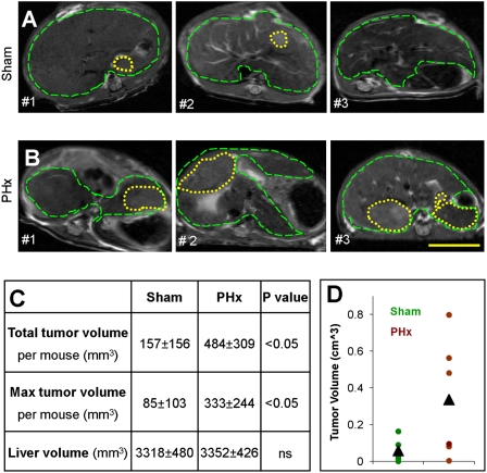
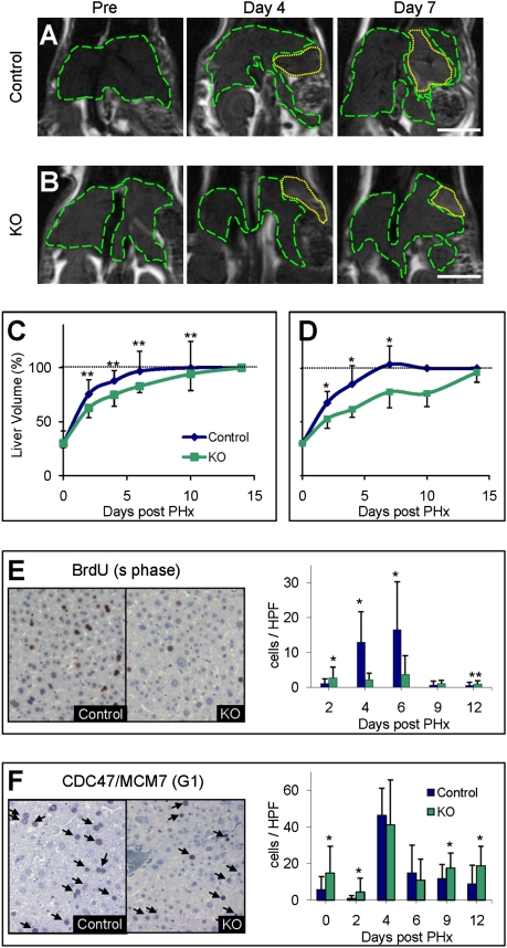
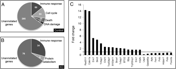
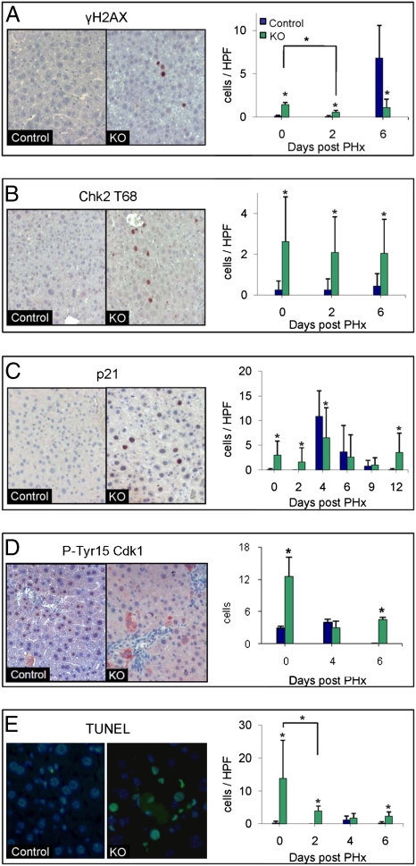
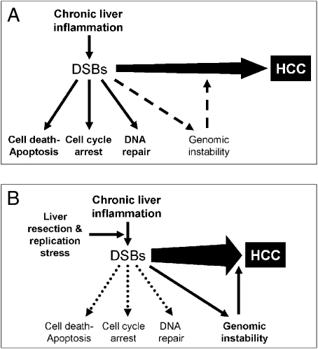
Hepatocellular carcinoma (HCC) is the third leading cause of cancer mortality worldwide and is considered to be the outcome of chronic liver inflammation. Currently, the main treatment for HCC is surgical resection. However, survival rates are suboptimal partially because of tumor recurrence in the remaining liver. Our aim was to understand the molecular mechanisms linking liver regeneration under chronic inflammation to hepatic tumorigenesis. Mdr2-KO mice, a model of inflammation-associated cancer, underwent partial hepatectomy (PHx), which led to enhanced hepatocarcinogenesis. Moreover, liver regeneration in these mice was severely attenuated. We demonstrate the activation of the DNA damage-response machinery and increased genomic instability during early liver inflammatory stages resulting in hepatocyte apoptosis, cell-cycle arrest, and senescence and suggest their involvement in tumor growth acceleration subsequent to PHx. We propose that under the regenerative proliferative stress induced by liver resection, the genomic unstable hepatocytes generated during chronic inflammation escape senescence and apoptosis and reenter the cell cycle, triggering the enhanced tumorigenesis. Thus, we clarify the immediate and long-term contributions of the DNA damage response to HCC development and recurrence.


Conflict of interest statement

The authors declare no conflict of interest.

Figures

Fig. 1.
Fig. 1.
Tumorigenesis acceleration. Representative T2-weighted axial MRI of three different Mdr2−/− mice per group aged 12 months that underwent sham surgery (A) or PHx (B) at the age of 9 months. Dashed green lines encircle the liver, and yellow dotted lines encircle tumors. (Scale bar: 1 cm.) (C) Table summing the tumor status at the age of 12 months of mice that underwent sham surgery (n = 5) or PHx (n = 10) at the age of 9 months, as assessed by MRI. (D) Liver tumor volume in Mdr2−/− mice operated on at the age of 3 months was assessed at the age of 12 months and plotted for each animal and for the group average of sham surgery (green, n = 5) or PHx (brown, n = 6) (P < 0.05).
Fig. 2.
Fig. 2.
Regenerative delay. Representative anatomical coronal MRI of the same 9-month-old control (A) and Mdr2−/− (KO) (B) mice before and on days 4 and 7 following PHx. Dashed green lines encircle the liver, and yellow dotted lines encircle the residue of the resected lobe. (Scale bar: 1 cm.) Liver volume monitoring after 70% PHx of 3-month-old (n = 8 per time point per group) (C) and 9-month-old (n ≥ 10 per time point per group) (D) in control (blue) and KO (green) mice. Percentage of liver volume compared with the volume before surgery was calculated for each animal, and averages ± SD were plotted. *P < 0.01; **P < 0.05. The horizontal dashed line marks liver volume restoration. Representative liver sections of control and KO 9-month-old mice killed on day 6 after 35% PHx immunostained for BrdU (E) and CDC47 (F). Quantification of the BrdU- and CDC47-positive cells (E and F, respectively) was done at the indicated time points for 30 randomly selected fields (n ≥ 3 mice per time point per group). HPF, high-power field.
Fig. 3.
Fig. 3.
Gene expression profile. Control or Mdr2−/− (KO) livers of 9-month-old mice that underwent PHx were analyzed by Affymetrix arrays. Functional analysis of the up-regulated genes on day 6 after PHx in control (A) or KO (B) mice. (C) Graph illustrating the fold change in gene expression of representative differentially regulated genes involved in DNA damage and repair between KO vs. control mice before PHx. The horizontal dashed line marks a fold change of 1 (no change).
Fig. 4.
Fig. 4.
Activation of DNA damage response. Representative liver sections of 9-month-old control and Mdr2−/− (KO) mice obtained before PHx. Slides were immunostained (red) for γ-H2AX (A), Chk2-T68 (B), P21 (C), and Tyr15-phosphorylated Cdk1 (D) and for apoptosis by TUNEL assay (E). (DAPI, blue; apoptosis, green). Quantification of immunostaining for all time points of γ-H2AX (A), Chk2-T68 (B), P21 (C) and p-Tyr15 Cdk1 (D) and for apoptosis (E) was done (n ≥ 30 per time point per group, *P < 0.001). HPF, high-power field.
Fig. 5.
Fig. 5.
Genomic instability. Proposed model to the enhanced tumorigenesis induced by liver resection under chronic inflammatory background. (A) Chronic liver inflammation induces many afflictions, including DSBs, by oxidative damage. All these ailments, together and apart, contribute to the progress of HCC. The DNA damage response leads to cell cycle arrest, DNA repair, and apoptosis (solid-line arrows). Accumulation of DNA damage results in genomic instability (dashed-line arrow). (B) While performing liver resection, we induced proliferative stress on the hepatocytes. Under the replicative stress, some of the impaired cells containing DSBs were salvaged from the DNA damage response and replicated, thus increasing genomic instability and facilitating tumor progression (solid-line arrow).

Similar articles

Cited by

References

    1. Karin M, Greten FR. NF-kappaB: Linking inflammation and immunity to cancer development and progression. Nat Rev Immunol. 2005;5:749–759. - PubMed
    1. Mantovani A, Allavena P, Sica A, Balkwill F. Cancer-related inflammation. Nature. 2008;454:436–444. - PubMed
    1. O’Byrne KJ, Dalgleish AG. Chronic immune activation and inflammation as the cause of malignancy. Br J Cancer. 2001;85:473–483. - PMC - PubMed
    1. Kundu JK, Surh YJ. Inflammation: Gearing the journey to cancer. Mutat Res. 2008;659:15–30. - PubMed
    1. Coussens LM, Werb Z. Inflammation and cancer. Nature. 2002;420:860–867. - PMC - PubMed

Publication types

MeSH terms

Substances

Associated data