. 2010 Feb 2;107(5):2247-52.
doi: 10.1073/pnas.0907908107. Epub 2010 Jan 19.

T7 phage protein Gp2 inhibits the Escherichia coli RNA polymerase by antagonizing stable DNA strand separation near the transcription start site

Affiliations

T7 phage protein Gp2 inhibits the Escherichia coli RNA polymerase by antagonizing stable DNA strand separation near the transcription start site

Beatriz Cámara et al. Proc Natl Acad Sci U S A. .

Abstract

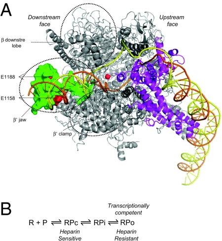
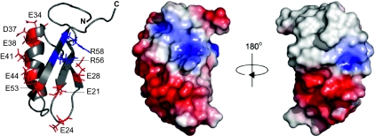
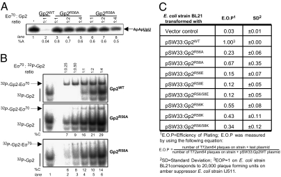
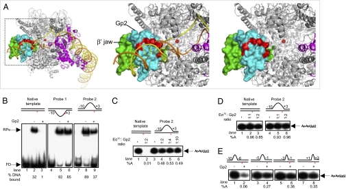
Infection of Escherichia coli by the T7 phage leads to rapid and selective inhibition of the host RNA polymerase (RNAP)--a multi-subunit enzyme responsible for gene transcription--by a small ( approximately 7 kDa) phage-encoded protein called Gp2. Gp2 is also a potent inhibitor of E. coli RNAP in vitro. Here we describe the first atomic resolution structure of Gp2, which reveals a distinct run of surface-exposed negatively charged amino acid residues on one side of the molecule. Our comprehensive mutagenesis data reveal that two conserved arginine residues located on the opposite side of Gp2 are important for binding to and inhibition of RNAP. Based on a structural model of the Gp2-RNAP complex, we propose that inhibition of transcription by Gp2 involves prevention of RNAP-promoter DNA interactions required for stable DNA strand separation and maintenance of the "transcription bubble" near the transcription start site, an obligatory step in the formation of a transcriptionally competent promoter complex.


Conflict of interest statement

The authors declare no conflict of interest.

Figures

Fig. 1.
Fig. 1.
(A) Model of the RPo generated using the crystal structure of Thermus aquaticus RNAP (25) shown as a ribbon representation. Highlighted in green is the β′ jaw domain at the “downstream face” of the RNAP (E. coli residues 1149–1190), the deletion of which confers Gp2 resistance. The locations of amino acids E1158 and E1188 are indicated in red. The active site is indicated by the orange sphere. The location of the σ factor (magenta) at the “upstream face” of the RNAP and the path of the modeled DNA (orange, template strand; yellow, nontemplate strand) in the RPo is shown. Circled are the domains of the β and β′ subunit at the downstream face of the RNAP, which together with the β′ jaw contribute to the downstream DNA-binding channel. (The β′ GNCD domain is absent in the T. aquaticus RNAP.) (B) Schematic depiction of the steps leading to the transcriptionally competent RPo at the lacUV5 promoter (12). R, RNAP; P, promoter template; RPc, closed promoter complex; RPi, intermediate promoter complex; RPo, open promoter complex.
Fig. 2.
Fig. 2.
NMR-derived three-dimensional structure of Gp2. The left panel shows a ribbon representation of Gp2 indicating the side chains of the negatively charged amino acid residues and the conserved arginines at positions 56 (red) and 58 (blue). The middle and right panels show two views of the molecular surface of Gp2 (in the same orientation as the ribbon form) color-coded according to a basic electrostatic surface distribution, calculated using the vacuum electrostatics program in Pymol, version 0.99rc6.
Fig. 3.
Fig. 3.
Role of R56 and R58 in Gp2 function. (A) An autoradiograph of a 20% (wt/vol) denaturing gel showing synthesis of the transcript ApApUpU (indicated by the arrow; with the underlined nucleotides 32P-labeled) from lacUV5 by Eσ70 in the presence of increasing amounts of Gp2R56A and Gp2R58A. The percentage ApApUpU synthesized (%A) by Eσ70 in the presence of Gp2 with respect to reactions with no Gp2 are given at the bottom of the gels. (B) An autoradiograph of a 4.5% (wt/vol) native gel showing the binding of 32P-Gp2WT (Top), 32P-Gp2R56A (Middle), and 32P-Gp2R58A (Bottom) to Eσ70 are shown. The migration positions of 32P-Gp2 (lane 1) and the Eσ70-Gp2 complex (lanes 2–6) are indicated. Radioactivity in the mutant and WT Eσ70-Gp2 complexes was measured, and the Eσ70-binding activity of the R56A and R58A Gp2 mutants is expressed as the percentage of Gp2WT-binding activity (%C) for each corresponding ratio of Eσ70:32P-Gp2. At the bottom of the middle and bottom panels, the percentage of mutant 32P-Gp2 associated with Eσ70 compared with Gp2WT is given (%C). In A and B, the molar ratio of Gp2 present with respect to Eσ70 in each lane is shown at the top. (C) Plating efficiency of T72 AM64 phage on E. coli strain BL21 transformed with pSW33gp2 encoding mutant Gp2 proteins with the R to E and R to K substitutions at positions 56 and/or 58.
Fig. 4.
Fig. 4.
Gp2 inhibits step(s) leading to the RPo. (A) Model of the Gp2–RNAP complex. The boxed region in the image in the left panel is enlarged in the middle (with promoter DNA) and right panels (without promoter DNA) to emphasize the β′ jaw region. The RNAP is presented as in Fig. 1A. Gp2 is shown in cyan, and the negatively charged side chains of residues E21, E28, E34, D37, E38, E41, E44 and E53, which protrude into the DNA binding channel, are highlighted in red. (B) Autoradiograph of a 4.5% (wt/vol) native gel showing heparin-resistant RPo formation by Eσ70 in the absence (lanes 2, 5, and 8) and presence (lanes 3, 6, and 9) of ∼2-fold molar excess of Gp2 on 32P-labeled versions of the fully duplex (native) and heteroduplex probes 1 and 2 lacUV5 promoter templates (see text; FD, free DNA). The % template DNA in the RPo is shown at the bottom of the gel. (C) Autoradiograph of a 20% (wt/vol) denaturing gel showing synthesis of the transcript ApApUpU (indicated by the arrow) from the native (lanes 1 and 2) and heteroduplex probe 2 (lanes 3–6) lacUV5 promoter templates in the absence (lanes 1 and 3) and presence (lanes 2, 4, 5, and 6) of Gp2. The Gp2:Eσ70 molar ratio in each lane is shown at the Top. The percentage transcripts synthesized (%A) by Eσ70 in the presence of Gp2 with respect to reactions with no Gp2 are given at the bottom. (D) As in C, but Gp2 was added to the reactions after transcriptionally competent promoter complexes had formed on native (lanes 2 and 3) and heteroduplex probe 2 (lanes 5 and 6) lacUV5 promoter templates. (E) As in C, but assays were conducted with lacUV5 promoter templates containing heteroduplex segments of different lengths and at different positions with respect to the transcription start site (probes 3–6; see text). In BE, the lacUV5 promoter templates used are shown schematically with the positions and lengths of the heteroduplex segment indicated with respect to +1 site (indicated by the red asterisk).

Similar articles

Cited by

References

    1. Haugen SP, Ross W, Gourse RL. Advances in bacterial promoter recognition and its control by factors that do not bind DNA. Nat Rev Microbiol. 2008;6:507–519. - PMC - PubMed
    1. Wigneshweraraj S, et al. Modus operandi of the bacterial RNA polymerase containing the sigma54 promoter-specificity factor. Mol Microbiol. 2008;68:538–546. - PubMed
    1. Nechaev S, Severinov K. The elusive object of desire—interactions of bacteriophages and their hosts. Curr Opin Microbiol. 2008;11:186–193. - PMC - PubMed
    1. Nechaev S, Severinov K. Bacteriophage-induced modifications of host RNA polymerase. Annu Rev Microbiol. 2003;57:301–322. - PubMed
    1. Hesselbach BA, Nakada D. “Host shutoff” function of bacteriophage T7: Involvement of T7 gene 2 and gene 0.7 in the inactivation of Escherichia coli RNA polymerase. J Virol. 1977;24:736–745. - PMC - PubMed

Publication types

MeSH terms

Associated data

LinkOut - more resources