. 2010 Feb 2;107(5):2283-8.
doi: 10.1073/pnas.0909434107. Epub 2010 Jan 19.

Functional reciprocity between Na+ channel Nav1.6 and beta1 subunits in the coordinated regulation of excitability and neurite outgrowth

Affiliations

Functional reciprocity between Na+ channel Nav1.6 and beta1 subunits in the coordinated regulation of excitability and neurite outgrowth

William J Brackenbury et al. Proc Natl Acad Sci U S A. .

Abstract

Voltage-gated Na(+) channel (VGSC) beta1 subunits regulate cell-cell adhesion and channel activity in vitro. We previously showed that beta1 promotes neurite outgrowth in cerebellar granule neurons (CGNs) via homophilic cell adhesion, fyn kinase, and contactin. Here we demonstrate that beta1-mediated neurite outgrowth requires Na(+) current (I(Na)) mediated by Na(v)1.6. In addition, beta1 is required for high-frequency action potential firing. Transient I(Na) is unchanged in Scn1b (beta1) null CGNs; however, the resurgent I(Na), thought to underlie high-frequency firing in Na(v)1.6-expressing cerebellar neurons, is reduced. The proportion of axon initial segments (AIS) expressing Na(v)1.6 is reduced in Scn1b null cerebellar neurons. In place of Na(v)1.6 at the AIS, we observed an increase in Na(v)1.1, whereas Na(v)1.2 was unchanged. This indicates that beta1 is required for normal localization of Na(v)1.6 at the AIS during the postnatal developmental switch to Na(v)1.6-mediated high-frequency firing. In agreement with this, beta1 is normally expressed with alpha subunits at the AIS of P14 CGNs. We propose reciprocity of function between beta1 and Na(v)1.6 such that beta1-mediated neurite outgrowth requires Na(v)1.6-mediated I(Na), and Na(v)1.6 localization and consequent high-frequency firing require beta1. We conclude that VGSC subunits function in macromolecular signaling complexes regulating both neuronal excitability and migration during cerebellar development.


Conflict of interest statement

The authors declare no conflict of interest.

Figures

Fig. 1.
Fig. 1.
β1-mediated neurite outgrowth is inhibited by TTX and the Scn8a null mutation. (A) Neurite length of WT CGNs grown on CHL or CHL-β1 monolayers and treated with/without TTX (10 μM) for 48 h (n = 20). (B) Neurite distribution (%) plotted against neurite length for CGNs in A. (C) Neurite length of WT CGNs grown on CHL monolayers and treated with/without TTX and/or FGF (20 ng/mL) for 48 h (n = 300). (D) Neurite distribution (%) plotted against neurite length for CGNs in C. (E) Neurite lengths of WT and Scn8a null CGNs grown on CHL or CHL-β1 monolayers (n = 300). (F) Neurite distribution (%) plotted against neurite length for CGNs in E. (G) Neurite lengths of WT and Scn8a null CGNs grown on CHL monolayers and treated with/without FGF for 48 h (n = 300). (H) Neurite distribution (%) plotted against neurite length for CGNs in G. Data are mean ± SEM. ***P < 0.001, ANOVA with Tukey’s post hoc test. (I) Western blot of WT and Scn8a null cerebellar membrane protein, using anti-β1. Anti-α-tubulin was used as a loading control.
Fig. 2.
Fig. 2.
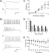
Electrical excitability is impaired in Scn1b null CGNs in vivo. (A) Voltage (V)-gated currents in a (i) WT and (ii) Scn1b null CGN following depolarization to 0 mV. (B) Membrane V recordings from (i) WT and (ii) Scn1b null CGNs following current (I) injection. Upper traces represent CGNs that sustained repetitive firing during 500 ms; lower traces represent CGNs that failed. (C) AP firing rate plotted as a function of I, normalized to AP threshold for WT (filled circles) and Scn1b null (open circles) CGNs. Data are mean ± SEM (n ≥ 15). *P < 0.05; **P < 0.01; ***P < 0.001, t test. (D) Distribution of APs across the 500-ms I injection for WT (dark bars) and Scn1b null (light bars) CGNs. For each CGN, the number of APs was counted in each 100-ms interval for the sweep giving maximum firing frequency. Data are mean ± SEM (n ≥ 16). P < 0.001 between WT and Scn1b null (two-way ANOVA). (E) Instantaneous firing frequency (Inst. freq., in Hz), calculated as the reciprocal of the interval between two adjacent APs, for WT (filled circles) and Scn1b null (open circles) CGNs, plotted for the first 10 AP intervals. Data are expressed as mean ± SEM (n ≥ 11). P < 0.001 between WT and Scn1b null (two-way ANOVA).
Fig. 3.
Fig. 3.
Resurgent Na+ current (INa) is reduced in Scn1b null CGNs cultured for 14 days in vitro (DIV). (A) Whole-cell INa from WT (black), and Scn1b null (gray) CGNs elicited by 60-ms depolarizing voltage (V) pulses to −40 mV normalized to peak transient I (%). Inset, I persisting for 50–55 ms after onset of depolarization. (B) Resurgent I from WT (black) and Scn1b null (gray) CGNs elicited by repolarization to −30 mV following a depolarizing pulse to +30 mV for 20 ms, normalized to peak transient I at +30 mV (%). (C) Persistent I measured as the mean I at 50–55 ms after onset of depolarization (Left) and resurgent I (Right). Data are presented as mean ± SEM (n ≥ 19). *P = 0.01 (t test). (D) Western blots of WT and Scn1b null cerebellar membrane protein using anti-β4 and anti-β2. Anti–α-tubulin was used as a loading control.
Fig. 4.
Fig. 4.
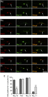
Nav1.6 expression is reduced at the AIS of Scn1b null CGNs. (AD) 100× Z-series confocal projections of WT (i) and Scn1b null (ii) 14 DIV CGNs labeled with anti-ankyrinG (AnkG) (red) and α subunit antibodies (green): Nav1.6 (A), pan-α subunit (B), Nav1.2 (C), and Nav1.1 (D). (Scale bar: 20 μm.) Arrows point to AIS expressing AnkG. (E) Proportion of AnkG-expressing CGN AIS that also express α subunits for WT (dark bars) and Scn1b null (light bars) (n = 60 fields of view taken from three mice of each genotype). Data are mean ± SEM. ***P <.001, Mann-Whitney test.
Fig. 5.
Fig. 5.
Nav1.6 expression is reduced at the AIS of Scn1b null Purkinje neurons. (A) 100× Z-series confocal projections of WT (i) and Scn1b null (ii) P14 cerebellum. AIS are labeled with anti-AnkG (red) and anti-Nav1.6 (green). Insets, 3× zoom of white boxes, showing colocalization of AnkG and Nav1.6 at Purkinje neuron AIS in WT, but not in Scn1b null. Arrows indicate Scn1b null Purkinje neuron AIS expressing both AnkG and Nav1.6. (B) Proportion of AnkG-expressing Purkinje neuron AIS that also express Nav1.6 for WT (dark bars) and Scn1b null (light bars) littermates (n = 60 fields of view from three mice of each genotype). Data are mean ± SEM. ***P < 0.001, t test. (C) 100× Z-series confocal projection of WT Purkinje neuron in situ labeled with anti-calbindin (red) and anti-β1 (green). The arrow shows β1 at AIS. (D) 100× Z-series confocal projection of WT 14 DIV CGN labeled with anti-pan VGSC α subunit (red) and anti-β1 (green). The arrow indicates β1 at AIS. (Scale bars: 20 μm.)
Fig. 6.
Fig. 6.
α subunits, β1, and contactin are present at the growth cone. 100× Z-series confocal projections of WT 14 DIV CGNs labeled with pan-α subunit (green), anti-β1 (A; red) or anti-contactin (B; red), and Alexa 594–conjugated phalloidin (magenta). (Scale bar: 10 μm.) he panels on the right show 4× digital zoom views highlighting growth cone (arrows). These distributions were observed in all three mice studied.
Fig. 7.
Fig. 7.
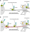
A model for INa involvement in β1-mediated neurite outgrowth. (A) In immature CGNs lacking AIS, complexes containing Nav1.6, β1, and contactin are present throughout the neuronal membrane in the soma, neurite, and growth cone. Localized Na+ influx is necessary for β1-mediated neurite extension and migration (11, 22, 25). VGSC complexes along the neurite are proposed to participate in cell–cell adhesion and fasciculation (22). (B) In 14 DIV CGNs, β1 is also required for Nav1.6 expression at the AIS, and subsequent high-frequency AP firing through modulation of resurgent INa (8, 10, 45). Electrical activity may further promote β1-mediated neurite outgrowth at or near the growth cone in vivo (8, 10, 45). Thus, the developmental functions of β1 and Nav1.6 are complementary, such that Na+ influx carried by Nav1.6 is required for β1-mediated neurite outgrowth and β1 is required for normal expression/activity of Nav1.6 at the AIS. Fyn kinase and AnkG also are likely present in all complexes (11), but they are only shown once in each panel for clarity. The FGF-mediated, β1-independent neurite outgrowth pathway is shown as well. Other CAMs that regulate neurite outgrowth and also may interact with β1 in this system (11, 26), have been omitted for clarity.

Similar articles

Cited by

References

    1. Catterall WA. From ionic currents to molecular mechanisms: The structure and function of voltage-gated sodium channels. Neuron. 2000;26:13–25. - PubMed
    1. Hille B. Ionic Channels of Excitable Membranes. 2nd Ed. Sunderland, MA: Sinauer Associates; 1992.
    1. Goldin AL, et al. Nomenclature of voltage-gated sodium channels. Neuron. 2000;28:365–368. - PubMed
    1. Trimmer JS, Rhodes KJ. Localization of voltage-gated ion channels in mammalian brain. Annu Rev Physiol. 2004;66:477–519. - PubMed
    1. Vacher H, Mohapatra DP, Trimmer JS. Localization and targeting of voltage-dependent ion channels in mammalian central neurons. Physiol Rev. 2008;88:1407–1447. - PMC - PubMed

Publication types

MeSH terms