. 2010 Mar 19;106(5):920-31.
doi: 10.1161/CIRCRESAHA.109.207274. Epub 2010 Feb 4.

Transmural flow modulates cell and fluid transport functions of lymphatic endothelium

Affiliations

Transmural flow modulates cell and fluid transport functions of lymphatic endothelium

Dimana O Miteva et al. Circ Res. .

Abstract

Rationale: Lymphatic transport of peripheral interstitial fluid and dendritic cells (DCs) is important for both adaptive immunity and maintenance of tolerance to self-antigens. Lymphatic drainage can change rapidly and dramatically on tissue injury or inflammation, and therefore increased fluid flow may serve as an important early cue for inflammation; however, the effects of transmural flow on lymphatic function are unknown.

Objective: Here we tested the hypothesis that lymph drainage regulates the fluid and cell transport functions of lymphatic endothelium.

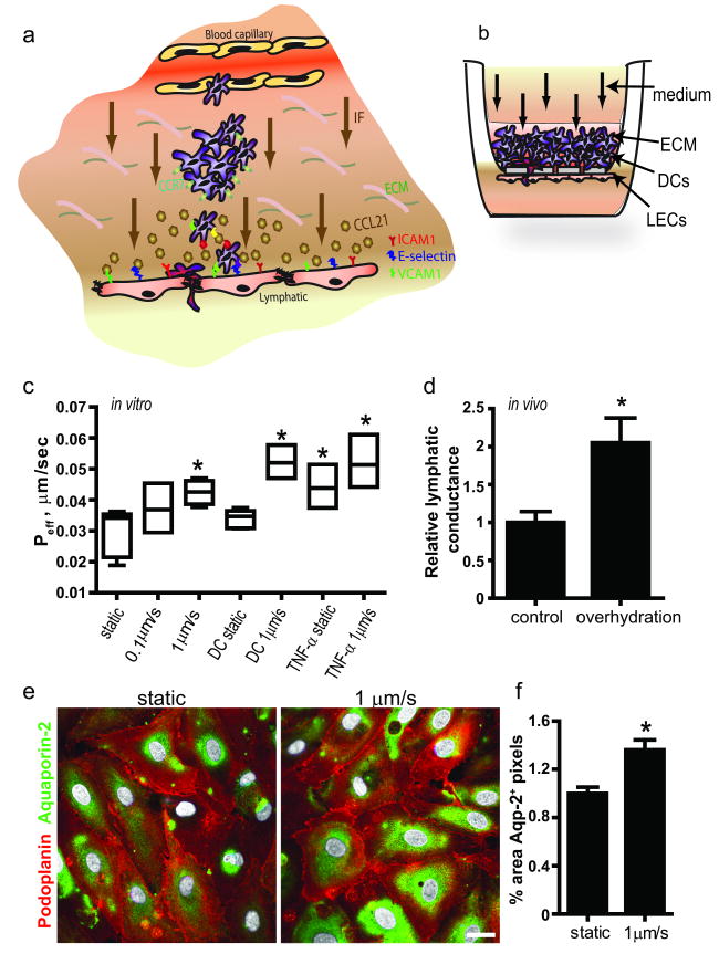
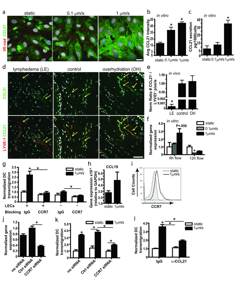
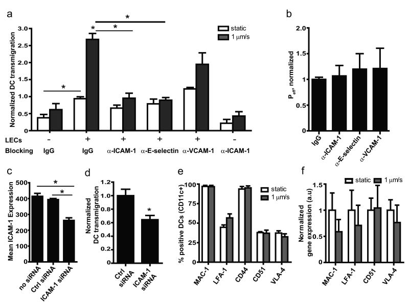
Methods and results: Using in vitro and in vivo models, we demonstrated that lymphatic endothelium is sensitive to low levels of transmural flow. Basal-to-luminal flow (0.1 and 1 mum/sec) increased lymphatic permeability, dextran transport, and aquaporin-2 expression, as well as DC transmigration into lymphatics. The latter was associated with increased lymphatic expression of the DC homing chemokine CCL21 and the adhesion molecules intercellular adhesion molecule-1 and E-selectin. In addition, transmural flow induced delocalization and downregulation of vascular endothelial cadherin and PECAM-1 (platelet/endothelial cell adhesion molecule-1). Flow-enhanced DC transmigration could be reversed by blocking CCR7, intercellular adhesion molecule-1, or E-selectin. In an experimental model of lymphedema, where lymphatic drainage is greatly reduced or absent, lymphatic endothelial expression of CCL21 was nearly absent.

Conclusions: These findings introduce transmural flow as an important regulator of lymphatic endothelial function and suggest that flow might serve as an early inflammatory signal for lymphatics, causing them to regulate transport functions to facilitate the delivery of soluble antigens and DCs to lymph nodes.


Conflict of interest statement

Disclosures. None.

Figures

Figure 1
Figure 1. Fluid transport across lymphatic endothelium is increased after transmural flow preconditioning
a, Schematic showing transmural flow and migration of dendritic cells (DCs) from the extracellular matrix (ECM) across lymphatic endothelium. CCR7+ DCs chemoattract towards CCL21-secreting lymphatic vessels and transmigrate using the adhesion molecules ICAM-1, E-selectin and VCAM-1. b, The in vitro model of the DC-lymphatic microenvironment includes lymphatic endothelial cells (LECs) seeded on the bottom of a porous culture insert and ECM containing DCs through which medium flows at 0.1 or 1μm/s, from the basal to the apical side. c, Effective permeability of LECs to 3 kDa dextran after 12 h preconditioning with transmural flow, DCs, or 100 ng/ml TNF-α. d, Lymphatic conductance (volume drained fluid per tissue volume per time and applied pressure gradient) is roughly doubled in overhydratated vs. control mice. e, Aquaporin-2 expression in vitro under static vs. flow conditions. Bar, 20μm. f, Aquaporin-2 protein expression is increased after 12h transmural flow. *P<0.05 compared to static controls.
Figure 2
Figure 2. Transmural flow downregulates and delocalizes VE-cadherin and PECAM-1 on lymphatic endothelium
a, Immunostaining for VE-cadherin (red) and PECAM-1 (green) with 3D reconstruction of lymphatic endothelial cell (LEC) junctions in vitro after 12 h. In static conditions, PECAM-1 appears on overlapping portions of LECs surrounded by VE-cadherin (yellow arrows); in flow conditions, PECAM-1 and VE-cadherin appear delocalized and downregulated, with VE-cadherin localizing to the basal side (lower right). Bar, 20 μm. b, Image quantification shows downregulation of VE-cadherin and PECAM-1 with 1 μm/s flow after 12 h. c, Quantitative PCR for VE-cadherin and PECAM-1 in LECs after 12h treatment with transmural flow or with dendritic cells (DCs). d and e, Colocalization of VE-cadherin and PECAM-1 in the (d) x-y plane and (e) z-plane. f, Relative distributions of VE-cadherin and PECAM-1 show that VE-cadherin becomes localized to the basal surface upon flow activation. g, Quantification of in vivo immunostaining for VE-cadherin and PECAM-1 (associated with LYVE-1+ lymphatic structures) after 24h overhydration (OH).
Figure 3
Figure 3. Dendritic cell transmigration across lymphatic endothelium is increased by transmural flow and occurs through both transcellular and paracellular routes
a, In vitro dendritic cell (DC) transmigration across cultured lymphatic endothelial cells (LECs) after 12 h transmural flow and 100ng/ml TNF-α treatment. b, In vivo DC migration to the draining lymph node (LN) 12 h after adoptive transfer in control vs. overhydrated mice. White and black circles represent data from BALB/c and C57BL/6 mice, respectively. c, Paracellular and d, transcellular transmigration routes of DCs were seen in vitro. Top row: Confocal images of DCs (green, CD11c) in the process of transmigration across LECs (violet, phalloidin; red, VE-cadherin). Right and bottom insets to the first row of images show cross-sections in the y- and x-directions, respectively, with dotted lines indicating the insert membranes. Bar, 10μm. Bottom row: scanning electron micrographs show DC migration through both paracellular (left) and transcellular (right) pathways. Bars 10 μm (left), 4 μm (right).
Figure 4
Figure 4. Transmural flow increases CCL21 secretion by lymphatic endothelium
a, Representative confocal images show CCL21 (green) expressed by lymphatic endothelial cells (LECs) after 12 h exposure to 0 (static), 0.1, and 1μm/sec flow; red, VE-cadherin; bar, 20μm. b, Image quantification of CCL21 protein in cells exposed to 12h flow vs. static conditions. c, CCL21 protein measured by ELISA after 24h culture. d, Representative images of lymphatic vessels (red, LYVE-1) and CCL21 (green) in lymphedematous (LE), control, and overhydrated (OH) skin. Arrows indicate lymphatic vessels; bar, 50μm. e, Quantification of CCL21 staining in cultured LECs after 12h transmural flow. f, Real-time PCR expression after 6h and 12h flow treatment. g, DC transmigration across a LEC monolayer with control IgG or anti-CCR7 blocking antibodies. h, CCL19 gene expression by DCs in 3D cultures after 12h static or flow conditions. i, Representative histogram from flow cytometry showing no differences in CCR7 expression by DCs in static (dotted line) and 12h flow (1μm/sec, solid line) conditions; shaded area shows the negative control. j, Real-time PCR for CCR7 expression in DCs after 24h of siRNA transfection. k, DC transmigration across a LEC monolayer following DC transfection with control or CCR7 siRNA. l, CCL21 blocking inhibits the flow-enhanced DC transmigration across LECs. *P<0.05 compared to static controls.
Figure 5
Figure 5. Lymphatic expression of adhesion molecules is regulated by transmural flow
a, Immunostaining for ICAM-1, E-selectin and VCAM-1 (green) on cultured lymphatic endothelial cells (LECs) after 12h flow. Bar, 20μm. b, Image quantification from cultured LECs. c, Quantification of ICAM-1+, E-selectin+, and VCAM-1+ pixels associate with lymphatic structures (LYVE-1+) in mouse tail skin sections. d, Normalized gene expression after 6h and 12h flow; black bars show 12h flow in the presence of mature DCs. e, Confocol z-slices after 12h of 1μm/s flow, podoplanin (Podo, red) delineates the cell membrane; bar, 5 μm. *P<0.05 compared to static.
Figure 6
Figure 6. ICAM and E-selectin regulate flow-enhanced, but not static, DC transmigration
a, Transmigration of dendritic cells (DCs) across lymphatic endothelial cells (LECs) in presence of blocking antibodies after 12h flow (black bars) or static (white bars) conditions. b, No change in effective permeability to dextran of LECs blocked with ICAM-1, E-selectin or VCAM-1 after 12h flow. c, ICAM-1 protein on LECs following ICAM-1 siRNA knockdown; cells were stimulated for 12h with 10ng/ml TNF-α before measurement. d, DC transmigration across ICAM-1-silenced LECs under 1μm/s flow conditions. e and f, DC surface expression by flow cytometry (e) or gene expression by real-time PCR (f) of receptors to ICAM-1, VCAM-1, and E-selectin are not affected by flow.

References

    1. Itano AA, Jenkins MK. Antigen presentation to naive CD4 T cells in the lymph node. Nature Immunol. 2003;4:733–739. - PubMed
    1. Pape KA, Catron DM, Itano AA, Jenkins MK. The humoral immune response is initiated in lymph nodes by B cells that acquire soluble antigen directly in the follicles. Immunity. 2007;26:491–502. - PubMed
    1. Randolph GJ, Angeli V, Swartz MA. Dendritic-cell trafficking to lymph nodes through lymphatic vessels. Nat Rev Immunol. 2005;5:617–628. - PubMed
    1. Förster R, Davalos-Misslitz A, Rot A. CCR7 and its ligands: balancing immunity and tolerance. Nat Rev Immunol. 2008;8:362–371. - PubMed
    1. Bates DO, Hillman NJ, Williams B, Neal CR, Pocock TM. Regulation of microvascular permeability by vascular endothelial growth factors. J Anat. 2002;200:581–597. - PMC - PubMed

Publication types

MeSH terms