. 2010 Apr 9;285(15):11617-27.
doi: 10.1074/jbc.M109.094961. Epub 2010 Feb 4.

Inhibiting TRAF2-mediated activation of NF-kappaB facilitates induction of AP-1

Affiliations

Inhibiting TRAF2-mediated activation of NF-kappaB facilitates induction of AP-1

Sunil K Manna et al. J Biol Chem. .

Abstract

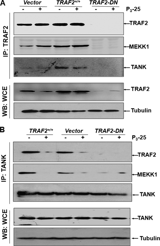
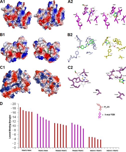
The compound 5-(4-methoxyarylimino)-2-N-(3,4-dichlorophenyl)-3-oxo-1,2,4-thiadiazolidine (P(3)-25) is known to possess anti-bacterial, anti-fungal, and anti-tubercular activities. In this report, we provide evidence that P(3)-25 inhibits NF-kappaB, known to induce inflammatory and tumorigenic responses. It activates AP-1, another transcription factor. It inhibits TRAF2-mediated NF-kappaB activation but not TRAF6-mediated NF-kappaB DNA binding by preventing its association with TANK (TRAF for NF-kappaB). It facilitates binding of MEKK1 with TRAF2 and thereby activates JNK and AP-1. We provide evidence, for the first time, that suggests that the interaction of P(3)-25 with TRAF2 leads to inhibition of the NF-kappaB pathway and activation of AP-1 pathway. These results suggest novel approaches to design of P(3)-25 as an anti-cancer/inflammatory drug for therapy through regulation of the TRAF2 pathway.


Figures

FIGURE 1.
FIGURE 1.
Effect of P3-25 on activation of NF-κB, AP-1, IKK, and JNK in Jurkat and HuT-78 cells. Jurkat and HuT-78 cells were treated with 100 nm P3-25 for different times, and nuclear extracts were prepared. NF-κB (A1) and AP-1 (B1) DNA binding was measured by a gel shift assay. Cells were co-transfected with plasmids for NF-κB promoter DNA that had been linked to SEAP (NFB-SEAP) or AP-1 promoter DNA that was linked to the luciferase reporter gene (AP-1-luciferase) and GFP. After washing, cells were cultured for 12 h and then treated with P3-25 (100 nm) for different times. The GFP-positive cells were counted, and transfection efficiency was calculated. Culture supernatant was assayed for SEAP activity. The results are represented as -fold activation over the nontransfected control (A2). After treatment, the cell pellet was extracted and assayed for luciferase activity (B2). Cells were treated with 100 nm P3-25 for different times. The whole cell extracts (300 μg of protein) were immunoprecipitated with anti-IKKα and IKKβ (1 μg each) antibodies, and kinase was assayed using GST-IκBα as a substrate (C). The IKKα was assayed from 100 μg of protein extract by Western blot. The amount of phospho-JNK was measured from 100 μg of whole cell extracts by Western blot using anti-phospho-JNK antibody (D, top). The whole cell extracts (300 μg of protein) were immunoprecipitated with 1 μg of anti-JNK antibody, and JNK activity was assayed using GST-Jun as substrate (D, bottom). HuT-78 cells, transfected with the NFB-SEAP construct were treated with 100 pm P3-25 or 5-aryl TZD for different times, and activity of SEAP was measured from culture supernatant (E).
FIGURE 2.
FIGURE 2.
Effect of P3-25 on TNF- or IL-8-induced NF-κB activation and PKAα activity. Jurkat cells were treated with 100 nm P3-25 for different times and then stimulated with 100 pm TNF or 100 ng/ml IL-8 for 2 h. NF-κB DNA binding was measured from nuclear extracts (A). The whole cell extracts (300 μg of protein), prepared from P3-25-treated but TNF- or IL-8-stimulated cells for 1 h, were immunoprecipitated with anti-PKAα (1 μg) antibody. PKAα activity was assayed using GST-p65 as substrate (C). The amount of PKAα was detected in the cell extract by Western blot. Jurkat cells were transfected with NFB-SEAP and GFP constructs cultured for 12 h and treated with P3-25 for different times. Cells were then stimulated with TNF (100 pm) or IL-8 (100 ng/ml) for 12 h. Culture supernatants were used to detect SEAP activity, and activity is indicated as -fold activation relative to vector-transfected cells (B).
FIGURE 3.
FIGURE 3.
Effect of P3-25 on TRAF2- and/or TRAF6-induced NF-κB, AP-1, IKK, and JNK activation. Jurkat cells were transfected with plasmids for NFB-SEAP or AP-1-luciferase and GFP. After washing, cells were cultured for 12 h and then treated with P3-25 (100 nm) for different times. Nuclear extracts were prepared, and NF-κB (A1) and AP-1 (B1) DNA binding was measured by gel shift assay. Culture supernatant was assayed for SEAP activity (A2). Cell pellet was extracted and assayed for luciferase activity (B2). The results are represented as -fold activation over the nontransfected control. TRAF2- and/or TRAF6-transfected cells were treated with P3-25 for different times. The whole cell extract (300 μg of protein) was immunoprecipitated with anti-IKKα and IKKβ (1 μg of each) antibodies, and IKK was assayed using GST-IκBα as substrate (C). The IKKα was assayed from 100 μg of protein extract by Western blot. The amounts of phospho-JNK were measured from 100 μg of whole cell extracts by Western blot (D, top). The whole cell extracts (300 μg of protein) were immunoprecipitated with anti-JNK antibody (1 μg), and JNK activity was measured using GST-Jun as substrate (D, bottom).
FIGURE 4.
FIGURE 4.
Effect of P3-25 on TRAF2, TANK, and MEKK1 interaction. HuT-78 cells were transfected with vector, TRAF2, or TRAF2-DN constructs. Cells were cultured for 12 h and then treated with P3-25 (100 nm) for 12 h. The whole cell extracts were prepared and incubated with Sepharose CL 4B beads coupled with anti-TRAF2 (A) or anti-TANK (B) antibody for 4 h. Beads were packed in a 2-ml column, washed with 0.2 m NaCl solution, and then eluted with 0.5 m NaCl solution. The fractions were run in 12% SDS-PAGE, and TRAF2, MEKK1, and TANK were detected by Western blot. IP, immunoprecipitation; WB, Western blot.
FIGURE 5.
FIGURE 5.
Effect of P3-25 on interaction with TRAF2 and TRAF6 in silico. Shown is a surface representation of TRAF2, colored on the basis of electrostatic potential (−57.293kbT/e to +57.293kbT/e, where kb, T, and e are the Boltzmann constant, temperature, and electron charge, respectively) and the bound P3-25. A surface representation of TRAF6 is shown with the bound P3-25 (A). P3-25 bound into the binding site of TRAF2, represented in stick form (light blue), forms hydrogen bonds with Arg393, Asp399, and Phe447 and the binding site of TRAF6 and forms hydrogen bonds with Arg392, Lys469, and Gly470 (B). The lowest energies of different clusters of TRAF2 and TRAF6 that bind with P3-25 have been calculated and are indicated in the graph (C).
FIGURE 6.
FIGURE 6.
Effect of P3-25 and 5-aryl TZD on interaction with TRADD-TRAF2, TRAF2-TANK, or TRAF2-MEKK complexes. A surface representation of TRADD-TRAF2 complex and binding of P3-25 and 5-aryl TZD at positive regions of the complex in silico is depicted (A1). Interaction of 5-aryl TZD and P3-25 with TRADD-TRAF2 complex is indicated (A2). Shown is a surface representation of the TRAF2-TANK complex, colored on the basis of electrostatic potential (−57.293kbT/e to +57.293kbT/e, where kb, T, and e are the Boltzmann constant, temperature and the electron charge, respectively) and the bound P3-25 and 5-aryl TZD (B1). P3-25 binds into the binding site of the TRAF2-TANK complex, represented in stick form (light blue), forms hydrogen bonds with Arg393, Asp399, and Phe447, and 5-aryl TZD forms hydrogen bonds with Arg393, Lys447, and Asn407 (B2). The electrostatic potential surface area of TRAF2-MEKK1 complex in binding with P3-25 and 5-aryl TZD is represented (C1). P3-25 interacts with the active site amino acid (magenta) Lys192 of MEKK1 and TRAF2 complex (C2, left). Interaction of 5-aryl TZD with MEKK1 active site residues (magenta) and TRAF2 complex is analyzed (C2, right). The graph shows the lowest binding energies of complexes with P3-25 and 5-aryl TZD with different complexes (D).
FIGURE 7.
FIGURE 7.
Schematic diagram of P3-25-mediated interference with cell signaling to inhibit NF-κB but to activate AP-1.

References

    1. Rayet B., Gelinas C. (1999) Oncogene 18, 6938–6947 - PubMed
    1. Karin M., Greten F. R. (2005) Nat. Rev. Immunol. 5, 749–759 - PubMed
    1. Baeuerle P. A., Baichwal V. R. (1997) Adv. Immunol. 65, 111–137 - PubMed
    1. Fujioka S., Niu J., Schmidt C., Sclabas G. M., Peng B., Uwagawa T., Li Z., Evans D. B., Abbruzzese J. L., Chiao P. J. (2004) Mol. Cell. Biol. 24, 7806–7819 - PMC - PubMed
    1. Wisdom R., Johnson R. S., Moore C. (1999) EMBO J. 18, 188–197 - PMC - PubMed

Publication types