. 2010 Apr 16;285(16):11937-47.
doi: 10.1074/jbc.M109.077933. Epub 2010 Feb 4.

Regulation of CD1 antigen-presenting complex stability

Affiliations

Regulation of CD1 antigen-presenting complex stability

Artur N Odyniec et al. J Biol Chem. .

Abstract

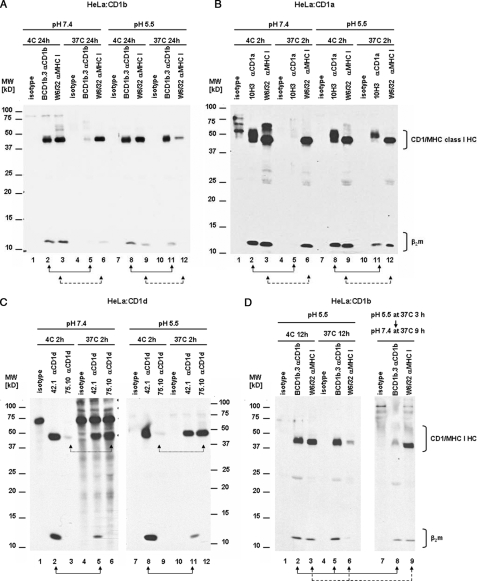
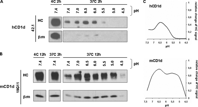
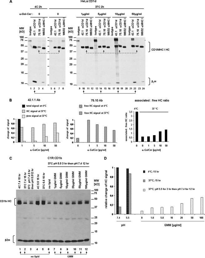
For major histocompatibility complex class I and II molecules, the binding of specific peptide antigens is essential for assembly and trafficking and is at the center of their quality control mechanism. However, the role of lipid antigen binding in stabilization and quality control of CD1 heavy chain (HC).beta(2)-microglobulin (beta(2)m) complexes is unclear. Furthermore, the distinct trafficking and loading routes of CD1 proteins take them from mildly acidic pH in early endososmal compartments (pH 6.0) to markedly acidic pH in lysosomes (pH 5.0) and back to neutral pH of the cell surface (pH 7.4). Here, we present evidence that the stability of each CD1 HC.beta(2)m complex is determined by the distinct pH optima identical to that of the intracellular compartments in which each CD1 isoform resides. Although stable at acidic endosomal pH, complexes are only stable at cell surface pH 7.4 when bound to specific lipid antigens. The proposed model outlines a quality control program that allows lipid exchange at low endosomal pH without dissociation of the CD1 HC.beta(2)m complex and then stabilizes the antigen-loaded complex at neutral pH at the cell surface.


Figures

FIGURE 1.
FIGURE 1.
CD1 HC·β2m complexes are unstable at 37 °C. CD1-transfected HeLa cells or C1R cells were surface bioyinylated, lysed with 0.5% Triton X-100 at pH 7.4, and lysates were incubated for 2–24 h at 4 or 37 °C. After immunoprecipitation with specific anti-CD1, anti-MHC class I or II, and anti-β2m antibodies, the immunoprecipitates were analyzed by SDS-PAGE (15%) under reducing conditions and immunoblotted with streptavidin-HRP. A, HeLa:CD1a cell lysates were incubated at 4 (lanes 1–4) or 37 °C (lanes 5–8) for 2 h. CD1a:HC·β2m complexes were immunoprecipitated with anti-CD1a 10H3 (lanes 2 and 6, solid line arrows), OKT6 (lanes 3 and 7, dotted line arrows), and 10D12 (lanes 4 and 8, dashed line arrows) antibodies. B, HeLa:CD1b cell lysates were incubated at 4 (lanes 1–3) or 37 °C (lanes 4–6) for 24 h. CD1b:HC·β2m complexes were immunoprecipitated with anti-CD1b BCD1.3 antibody (lanes 2 and 5) and MHC class I with W6/32 antibody (lanes 3 and 6). As a control, mouse IgG1 was used (isotype). C, C1R:CD1b cell lysates were incubated at 4 (lanes 1–5) or 37 °C (lanes 6–10) for 12 h. CD1b:HC·β2m complexes were immunoprecipitated with anti-CD1b BCD1b.3 (lanes 2 and 7, solid line arrows), anti-CD1b BCD1b.1 (lanes 3 and 8, dotted line arrows) antibodies, anti-β2m antibody BBM.1 (lanes 4 and 9, dashed line arrows), and MHC class II with anti-MHC II antibody L243 (lanes 5 and 10). As a control mouse IgG1 was used (isotype). D, HeLa:CD1d cell lysates were incubated at 4 (lanes 1–5) or 37 °C (lanes 6–10) for 2 h. CD1d HC·β2m complexes were immunoprecipitated with anti-CD1d 42.1 (lanes 2 and 7, solid line arrow) or 55.10 antibodies (lanes 3 and 8, dashed line arrows); free CD1d HC was immunoprecipitated with 75.10 antibody (lanes 4 and 9, dotted line arrows) and MHC class I with antibody W6/32 (lanes 5 and 10). E, C1R:CD1d cell lysates were incubated at 4 (lanes 1–5) or 37 °C (lanes 6–10) for 2 h. CD1d HC·β2m complexes were immunoprecipitated with anti-CD1d 42.1 (lanes 2 and 7, solid line arrows) or anti-β2m BBM.1 antibodies (lanes 4 and 9, dashed line arrows); free CD1d HC with anti-CD1d antibody 75.10 (lanes 4 and 9, dotted line arrows) and MHC class II with anti-MHC II L243 antibody (lanes 5 and 10). In all experiments mouse IgG2a or IgG1 were used as isotype controls. Arrows indicate the lanes to compare.
FIGURE 2.
FIGURE 2.
Rank order of CD1 isoform stability: CD1b > CD1c > CD1d > CD1a. HeLa:CD1a, CD1b, CD1c, or CD1d transfectants were surface biotinylated, lysed with 0.5% Triton X-100 (pH 7.4), and then lysates were incubated for 2 h at 4 or 37 °C. CD1 heavy chain/β2m complexes were immunoprecipitated with 10H3 (anti-CD1a, A), BCD1b.3 (anti-CD1b, B), F10/23.A.1 (anti-CD1c, C), or 42.1 (anti-CD1d, D) antibodies, whereas MHC class I:HC·β2m complexes were precipitated with antibody W6/32 (A–D). Mouse IgG1 and IgG2a were used as isotype controls. Immunoprecipitates were analyzed by SDS-PAGE (15%) in reducing conditions and immunoblotted with streptavidin-HRP. Arrows indicate lanes to compare. Quantification of band intensity was measured by densitometry using ImageJ software and results were calculated as proportion of HC or β2m signals at 37 °C compared with HC or β2m signals at 4 °C (A–D).
FIGURE 3.
FIGURE 3.
Stabilization of CD1a and -b HC·β2m complexes by low pH. CD1-transfected HeLa cells were surface biotinylated and lysed with 0.5% Triton X-100 (pH 7.4 or 5.5). Then lysates were incubated for 2–24 h at 4 or 37 °C. After immunoprecipitation with specific anti-CD1 and anti-MHC class I antibodies, immunoprecipitates were analyzed by SDS-PAGE (15%) in reducing conditions and immunoblotted with streptavidin-HRP. A, HeLa:CD1b cell lysates at pH 7.4 (lanes 1–6) or 5.5 (lanes 7–12) were incubated at 4 (lanes 1–3 and 7–9) or 37 °C (lanes 4–6 and 10–12) for 24 h. CD1b:HC·β2m complexes were immunoprecipitated with BCD1b.3 (lanes 2, 5, 8, and 11, solid line arrows) and MHC class I:HC·β2m complexes with W6/32 antibodies (lanes 3, 6, 9, and 12, dashed line arrows). B, HeLa:CD1a cell lysates at pH 7.4 (lanes 1–6) or 5.5 (lanes 7–12) were incubated at 4 (lanes 1–3 and 7–9) or 37 °C (lanes 4–6 and 10–12) for 2 h. CD1a:HC·β2m complexes were immunoprecipitated with 10H3 (lanes 2, 5, 8, and 11, solid lines arrows) and MHC class I:HC·β2m complexes with W6/32 antibodies (lanes 3, 6, 9, and 12, dashed line arrows). C, HeLa:CD1d cell lysates at pH 7.4 (lanes 1–6) or 5.5 (lanes 7–12) were incubated at 4 °C (lanes 1–3 and 7–9) or 37 °C (lanes 4–6 and 10–12) for 2 h. CD1d:HC·β2m complexes were immunoprecipitated with mAb 42.10 (lanes 2, 5, 8, and 11, solid line arrows) and the free CD1d HC was detected with mAb 75.10 (lanes 3, 6, 9, and 12, dotted line arrows). D, HeLa:CD1b cell lysates at pH 5.5 were incubated at 4 °C for 12 h (lanes 1–3) or 37 °C for 12 (lanes 4–6) or 3 h followed by pH neutralization to pH 7.4 with 1.5 m Tris-HCl buffer (pH 8.8) and a further 9-h incubation at 37 °C (lanes 7–9). CD1b HC·β2m complexes were immunoprecipitated with mAb BCD1b.3 (lanes 2, 5, and 8, solid line arrows) and MHC class I with mAb W6/32 (lanes 3, 6, and 9, dashed line arrows). In all experiments isotype-matched mouse IgG2a or IgG1 were used as controls. Arrows indicate lanes to compare.
FIGURE 4.
FIGURE 4.
pH-dependent protection of CD1 molecules corresponds to their locations in intracellular compartments. HeLa:CD1a, -CD1b, -CD1c, and -CD1d transfectants were surface biotinylated and lysed with 0.5% Triton X-100 at pH 7.4, 7.0, 6.5, 6.0, 5.5, 5.0, or 4.5. Lysates were incubated for 2 (CD1a and CD1d), 12 (CD1c), or 24 h (CD1b) at 37 °C. CD1 heavy chain·β2m complexes were immunoprecipitated with 10H3 (CD1a), BCD1b.3 (CD1b), F10/23.A.1 (CD1c), 42.1 or 75.10 (CD1d) antibodies. Immunoprecipitates were analyzed by SDS-PAGE (15%) in reducing conditions and immunoblotted with streptavidin-HRP. A, immunoprecipitated CD1 HC and β2m from lysates incubated at 37 °C at the indicated pH. B, quantification of band intensities for particular CD1 HC at different pH values by densitometry using ImageJ software. Curves indicate the ratio of signal for CD1 HC in lysates incubated at 37 °C at different pH in comparison to the incubation at 4 °C. C, the schematic shows the physiological pH of different intracellular compartments (ovals; ERC, endocytic recycling compartment; EE, early endosomes; LE, late endosomes), the intracellular location of CD1a, CD1b, and CD1c (black bars), and maximal protective effect of pH on the stability of CD1:HC·β2m complexes (gray triangles).
FIGURE 5.
FIGURE 5.
Effect of low pH on stability of human and mouse CD1d. HeLa:hCD1d and RAW:mCD1d transfectants were surface biotinylated and lysed with 0.5% Triton X-100 at pH 7.4, 7.0, 6.5, 6.0, 5.5, 5.0, or 4.5. Lysates were incubated for 2 h (hCD1d) or 2, 6, and 12 h (mCD1d) at 4 or 37 °C. Human CD1d:HC·β2m complexes were immunoprecipitated with anti-CD1d mAb 42.1 and 75.10, whereas mouse CD1d:HC·β2m complexes were immunoprecipitated with antibody 19G11. Immunoprecipitates were analyzed by SDS-PAGE (15%) in reducing conditions and immunoblotted with streptavidin-HRP. A and B, immunoprecipitated hCD1d and mCD1d HC and β2m from lysates incubated at 37 °C at the indicated pH. C, quantification of CD1d HC band intensities at different pH values by densitometry using ImageJ software. Curves indicate the ratio of signal for CD1d HC in lysates incubated at 37 °C at different pH values in comparison to the incubation at 4 °C.
FIGURE 6.
FIGURE 6.
Specific lipid antigens protect CD1d and CD1b HC·β2m complexes from disassembly. A, CD1d-transfected HeLa cells were surface biotinylated and lysed with 0.5% Triton X-100 (pH 7.4). Lysates were incubated for 2 h at 4 (lanes 1–4) or 37 °C (lanes 5–24) with different concentrations of α-GalCer (lanes 9–24) or without lipid (lanes 1–8). CD1d HC·β2m complexes were immunoprecipitated with anti-CD1d mAb 42.1 (lanes 2, 6, 10, 14, 18, and 22; solid line arrows), free CD1d HC were precipitated with mAb 75.10 (lanes 3, 7, 11, 15, 19, and 23; dashed line arrows), and MHC class I with antibody W6/32 (lanes 4, 8, 12, 16, 20, and 24). Mouse IgG1 and IgG2a were used as isotype controls. Immunoprecipitates were analyzed by SDS-PAGE (15%) under reducing conditions and immunoblotted with streptavidin-HRP. B, quantification of intensities of CD1d HC bands immunoprecipitated with 42.1 and 75.10 antibodies. Bars indicate the relative change of CD1d HC signal in samples incubated for 2 h at 37 °C in the presence of α-GalCer compared with incubation at 4 °C without lipid. Right graph shows the ratio between the associated and free form of CD1d HC in lysates incubated with increasing concentrations of α-GalCer at 37 or 4 °C without lipid. All measures were done by densitometry with ImageJ software. C, CD1b-transfected C1R cells were surface biotinylated and lysed with 0.5% Triton X-100 (pH 7.4) (lanes 1–3) or pH 5.5 (lanes 4–12). C32 GMM was added in increasing concentrations to samples run in lanes 7–12. Lysates were incubated for 15 h at 4 °C (lane 1 and 4), 15 h at 37 °C (lane 2 and 5), or 3 h at 37 °C followed by change in pH to 5.5 (lane 3) or 7.4 (lanes 6–12) and an additional 12-h incubation at the same temperature. CD1d HC·β2m complexes were immunoprecipitated with anti-β2m mAb BBM.1. Immunoprecipitates were analyzed by SDS-PAGE (15%) under reducing conditions and immunoblotted with streptavidin-HRP. D, quantification of intensities of bands corresponding to CD1b HC immunoprecipitated with BBM.1 antibody. Bars indicate the relative change of CD1d HC signal in samples incubated at different conditions (different temperatures and pH, in presence or absence of C32 GMM) compared with incubation at 4 °C without lipid. Intensities of bands were measured by densitometry with ImageJ software and relative change of signal was calculated as proportion of CD1b HC signal in samples incubated at 37 °C with or without GMM to intensity of CD1b HC signal at 4 °C.
FIGURE 7.
FIGURE 7.
Regulation of CD1 HC·β2m complex stability and quality control. A, mechanisms of the CD1 stability. At neutral pH in the absence of lipid antigen the CD1 HC·β2m complex is unstable (upper panel). At endosomal (acidic) pH, the CD1 HC·β2m complex is relatively more stable (middle panel). At endosomal pH, lipid antigen binding or exchange is facilitated, resulting in formation of the CD1 HC·β2m/lipid complex that is very stable even in neutral pH (lower panel). B, quality control model based on regulation of CD1 HC·β2m complex stability. In endosomal compartments, empty CD1 molecules are stabilized by low pH providing a pool of molecules available for binding lipid antigen. CD1 molecules loaded with proper lipid antigens in endosomal compartments form stable HC·β2m/lipid complexes that can traffic to the cell surface and remain stable at neutral pH. On the other hand, CD1 molecules that are empty or loaded with poor quality lipid are prone to dissociate at neutral pH, which would result in a shortened half-life at the cell surface. Relatively unstable complexes are depicted in blue and stable complexes are black.

Similar articles

Cited by

References

    1. Cohen N. R., Garg S., Brenner M. B. (2009) Adv. Immunol. 102, 1–94 - PubMed
    1. Kang S. J., Cresswell P. (2002) J. Biol. Chem. 277, 44838–44844 - PubMed
    1. De Silva A. D., Park J. J., Matsuki N., Stanic A. K., Brutkiewicz R. R., Medof M. E., Joyce S. (2002) J. Immunol. 168, 723–733 - PubMed
    1. Sugita M., Grant E. P., van Donselaar E., Hsu V. W., Rogers R. A., Peters P. J., Brenner M. B. (1999) Immunity 11, 743–752 - PubMed
    1. Barral D. C., Cavallari M., McCormick P. J., Garg S., Magee A. I., Bonifacino J. S., De Libero G., Brenner M. B. (2008) Traffic 9, 1446–1457 - PMC - PubMed

Publication types

MeSH terms