. 2010 Apr;298(4):G551-62.
doi: 10.1152/ajpgi.00497.2009. Epub 2010 Feb 4.

Insulin-like growth factors are more effective than progastrin in reversing proapoptotic effects of curcumin: critical role of p38MAPK

Affiliations

Insulin-like growth factors are more effective than progastrin in reversing proapoptotic effects of curcumin: critical role of p38MAPK

Pomila Singh et al. Am J Physiol Gastrointest Liver Physiol. 2010 Apr.

Abstract

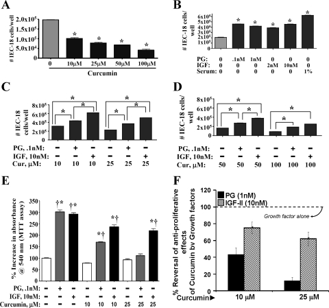
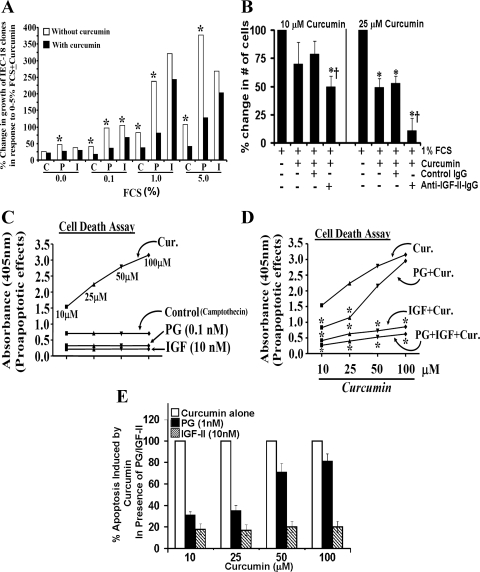
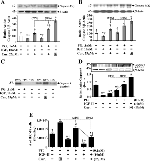
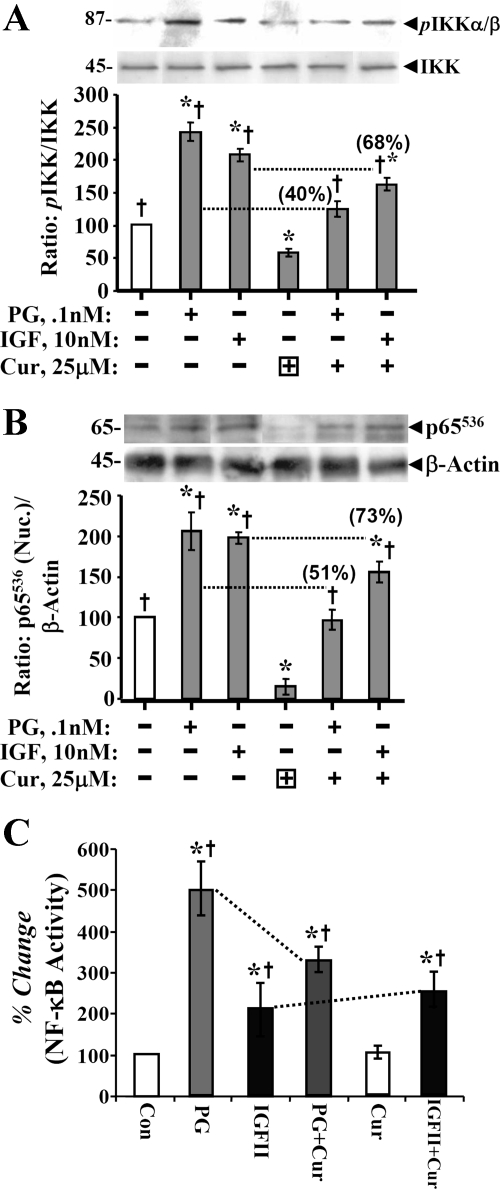
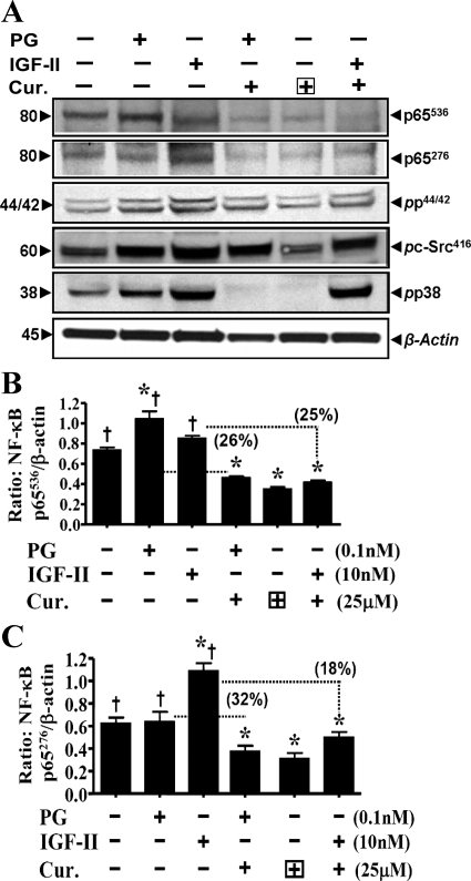
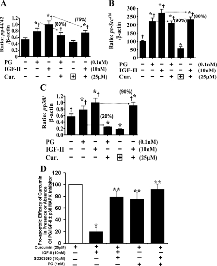
Progastrin and insulin-like growth factors (IGFs) stimulate hyperproliferation of intestinal epithelial cells (IECs) via endocrine/paracrine routes; hyperproliferation is a known risk factor for colon carcinogenesis. In the present study, inhibitory potency of curcumin in the presence or absence of progastrin and/or IGF-II was examined. Progastrin and IGF-II significantly increased proliferation of an immortalized IEC cell line, IEC-18, whereas curcumin decreased the proliferation in a dose-dependent manner. IGF-II was significantly more effective than progastrin in reversing antiproliferative effects of curcumin and reversed proapoptotic effects of curcumin by >80%; progastrin was relatively ineffective toward reversing proapoptotic effects of curcumin. IEC-18 clones were generated to overexpress either progastrin (IEC-PG) or hIGF-II (IEC-IGF). Proliferation of IEC-PG and IEC-IGF clones was increased, compared with that of control clones. Curcumin significantly reduced proliferation of IEC-PG, but not IEC-IGF, clones. Similarly, a human colon cancer cell line, Caco-2 (which expresses autocrine IGF-II), was relatively resistant to inhibitory effects of curcumin. However, Caco-2 cells treated with anti-IGF-II-antibodies were rendered sensitive to inhibitory effects of curcumin. Significant differences in inhibitory potency of curcumin against PG- vs. IGF-II-stimulated growth of IEC-18 cells were not reflected by differences in curcumin-mediated inhibition of activated (phosphorylated) ERKs/IKK(alpha/beta)/p65NF-kappaB and c-Src in wild-type (wt)IEC-18 cells, in response to the two growth factors. Surprisingly, curcumin was almost ineffective in reducing IGF-II-stimulated activation of p38MAPK but significantly reduced progastrin-stimulated phosphorylation of p38. Treatment with a p38MAPK inhibitor resulted in loss of protective effects of IGF-II against inhibitory effects of curcumin. These novel findings suggest that growth factor profile of patients and tumors may dictate inhibitory potency of curcumin and that combination of curcumin + p38MAPK inhibitor may be required for reducing hyperproliferative or tumorigenic response of IECs to endocrine and autocrine IGFs.


Figures

Fig. 1.
Fig. 1.
Effect of curcumin (Cur) ± IGF-II or progastrin (PG) on growth of IEC-18 cells (cell count assay). IEC-18 cells were treated with curcumin ± IGF-II or PG as described in materials and methods, and the effect was assessed in a cell count assay. Data are presented as means ± SE of 4 values from a representative of 3 experiments. *P < 0.05 vs. control values (no treatment) in A and B; *P < 0.05 vs. the indicated bars in C and D. E: effect of curcumin ± GF-II/PG on growth of IEC-18 cells (MTT assay). Absorbance measured in control wells (no treatment) was arbitrarily assigned a 100% value. Readings from all other wells are expressed as a % change from control values. Each bar represents mean ± SE of data from 6–8 wells from a representative of 2 experiments. *P < 0.05 vs. control; †P < 0.05 vs. curcumin alone. F: percent reversal of antiproliferative effects of curcumin, in the presence of growth factors. The % reversal of antiproliferative effects of curcumin by growth factors in an MTT assay (E), to levels measured in the presence of growth factors alone are shown. Values measured in the presence of growth factors alone were arbitrarily assigned 100%. Values presented for growth factor + curcumin are presented as a % of growth factor alone. *P < 0.05 vs. the corresponding values for PG + curcumin.
Fig. 2.
Fig. 2.
A: percent change in growth of IEC-18 clones in response to 0–5% FCS. IEC-18 clones, overexpressing either hPG (P) or hIGF-II (I), were used. Clones expressing vector alone served as control (C). Data (mean values) are derived from Table 1. Open bars, growth in response to fetal calf serum (FCS); solid bars, growth in response to FCS + curcumin (25 μM). Growth of control clones in response to 5% FCS was arbitrarily assigned 100% value; growth of all other clones is presented as a % of it. *P < 0.5 vs. filled (+curcumin) values. B: effect of anti-IGF-II-antibody treatment on the sensitivity of Caco-2 cells to inhibitory effects of curcumin. Caco-2 cells were plated in 35-mm cell culture dishes, as described previously (36). Since Caco-2 cells are extremely sensitive to loss of growth factors, the cells were maintained in 1% FCS from day 5 onward. On day 5 of cell culture, Caco-2 cells were treated with or without 10–25 μM (as shown) for 48 h, in the presence or absence of either the control (nonimmune) or specific anti-IGF-II-IgG (1:1,000 dilution). In each case the IgG was added 2 h before the addition of curcumin. At the end of the treatment, total number of cells/dish was counted by a cell count assay, as described under materials and methods. The % change in the number of cells is shown, wherein the total number of cells in the absence of curcumin treatment was arbitrarily assigned a 100% value. Each bar is mean ± SE of 6 dishes/2 experiments. *P < 0.05 vs. control (non-curcumin-treated values). †P < 0.05 vs. the corresponding control IgG values (3rd vs. 4th bar for the indicated curcumin-treatment). C and D: apoptotic death of wt IEC-18 cells (in a cell death assay) in response to camptothecin (control), in presence of curcumin ± growth factors. Apoptotic death was measured in terms of absorbance at 405 nm (y-axis). Increasing absorbance reflects increasing death. In C, absorbance of control or PG/IGF-II treated cells, are presented as flat lines, in relation to absorbance of curcumin-treated cells. In D, absorbance of samples treated with curcumin ± PG/IGF-II is presented. Each point = mean of 3 values from a representative of 3 experiments. Variation between values was ∼5–10%. *P < 0.05 vs. the corresponding value for curcumin alone. E: % apoptosis of cells induced by curcumin, in the presence of PG/IGF-II. Data presented are based on the data shown in D. Values measured in the presence of indicated concentration of curcumin alone were arbitrarily assigned 100% (open bar); apoptosis in the presence of factor alone was assigned 0%. Values presented for growth factor + curcumin are a % of cell death measured in response to curcumin alone. All values for IGF-II + curcumin-treated samples were significantly different (P < 0.05) from PG + curcumin-treated values.
Fig. 3.
Fig. 3.
IGF-II is more effective than PG in reversing proapoptotic effects of curcumin on IEC-18 cells. Proapoptotic effects of curcumin were measured in terms of either activation of caspase 3 (A and B) or caspase 9 (C and D), or number of floating cells (E). Representative Western blots (of 4 blots from 2 experiments) are presented in top panels. Relative levels of β-actin in the samples are shown. Ratio of activated caspases 3 or 9/β-actin was calculated, and control (nontreated) ratios were assigned 100% value. Ratios for all other treatments are presented as % of control values. In E, each bar = mean ± SE of floating cells, measured in six culture dishes/treatment. *P < 0.05 vs. control values; †P < 0.5 vs. curcumin alone. Horizontal dotted lines = curcumin-alone values. Dropdown vertical line = loss in proapoptotic effects in the presence of growth factor; % reversal of proapoptotic effects is presented numerically in parentheses on top of the indicated bar.
Fig. 4.
Fig. 4.
Inhibitory effect of curcumin on phosphorylation and activation of kinases in response to growth factors. A and B: phosphorylated kinase values for IKKα/β and pp65 NF-κB p65536 were calculated as ratio of total kinase/β-actin. Ratios of control (untreated) samples were assigned 100% value, and ratios of all other samples were calculated as a % of control. Data in each bar = means ± SE of 4 measurements from 2 experiments. Horizontal dotted lines = loss in levels of activated kinase in the presence of curcumin. The % loss of kinase activation, in response to IGF/PG, in the presence of curcumin is presented numerically in parentheses on top of the relevant bar. C: relative levels of activated NF-κB from a DNA binding assay. Each bar graph = mean ± SE of 3 measurements from 2 experiments. *P < 0.05 vs. control (untreated) samples; †P < 0.05 vs. curcumin alone.
Fig. 5.
Fig. 5.
A: representative Western blots for activated kinases in response to curcumin ± PG/IGF-II. Data presented is from one of 4 blots from 2 separate experiments. Relative levels of β-actin measured in a representative blot from corresponding samples is shown. B and C: inhibitory effects of curcumin on activation of pNF-κBp65536 (B) and, pNF-κBp65276 (C) in response to growth factors. Western blot data, as shown in A, were used to calculated the ratios as described in Fig. 4. Briefly, phosphorylated kinase values were calculated at a ratio of β-actin. Ratios of control (untreated) samples were assigned 100% values, and ratios of all other samples were calculated as a % of control. Data in each bar = means ± SE of 4 measurements from 2 experiments. Dotted horizontal lines = inhibitory effects of curcumin on activation of indicated kinases, as described in Fig. 4. Percent activation of kinase by PG/IGF-II was assigned 100% values; numerical values in parentheses = % loss in kinase activation in the presence of curcumin, compared with that in the presence of PG/IGF-II alone. *P < 0.05 vs. control; †P < 0.05 vs. curcumin alone.
Fig. 6.
Fig. 6.
AC: inhibitory effects of curcumin and activation of ERKs (A), c-Src (B), and p38MAPK (C), in response to growth factors. Western blot data, as shown in Fig. 5A, was used to calculate the ratios as described above for Fig. 5, B and C. Numerical values in parentheses are % loss in kinase activation in the presence of curcumin, compared with that in the presence of PG/IGF-II alone. *P < 0.05 vs. control; †P < 0.05 vs. curcumin alone. D: proapoptotic efficacy of curcumin in the presence or absence of PG/IGF-II ± p38MAPK inhibitor (measured in a cell death assay). On a scale of 0–100, cell death measured in the presence of curcumin alone was arbitrarily assigned 100%, whereas cell death in wild-type IEC-18 cells in the presence of growth factors alone was assigned 0%. Data represent means ± SE of 8 separate measurements from 2 experiments. *P < 0.05 vs. corresponding growth factor + curcumin values. p38 inhibitor (p38-I) (SB203580, 10 μM) was added 30 min before addition of growth factor ± curcumin, as previously described (32). Addition of p38 inhibitor alone did not significantly change levels of cell death measured (data not shown). *P < 0.05 vs. curcumin alone; **P < 0.05 vs. IGF-II + curcumin (second bar).

Similar articles

Cited by

References

    1. Barry J, Fritz M, Brender JR, Smith PE, Lee DK, Ramamoorthy A. Determining the effects of lipophilic drugs on membrane structure by solid-state NMR spectroscopy: the case of the antioxidant curcumin. J Am Chem Soc 131: 4490–44988, 2009 - PMC - PubMed
    1. Beevers CS, Chen L, Liu L, Luo Y, Webster NJ, Huang S. Curcumin disrupts the Mammalian target of rapamycin-raptor complex. Cancer Res 69: 1000–1008, 2009 - PMC - PubMed
    1. Bordonaro M, Mariadason JM, Aslam F, Heerdt BG, Augenlicht LH. Butyrate-induced apoptotic cascade in colonic carcinoma cells: modulation of the beta-catenin-Tcf pathway and concordance with effects of sulindac and trichostatin A, but not curcumin. Cell Growth Differ 10: 713–720, 1999 - PubMed
    1. Ciccotosto GD, McLeish A, Hardy KJ, Shulkes A. Expression, processing, and secretion of gastrin in patients with colorectal carcinoma. Gastroenterology 109: 1142–1153, 1995 - PubMed
    1. Cobb S, Wood T, Ceci J, Varro A, Velasco M, Singh P. Intestinal expression of progastrin significantly increases colon carcinogenesis in transgenic mice in response to AOM. Cancer 100: 1311–1323, 2004 - PubMed

Publication types

MeSH terms

Substances

LinkOut - more resources