. 2010 Feb;82(2):257-65.
doi: 10.4269/ajtmh.2010.09-0309.

Development of Cryptosporidium parvum-induced gastrointestinal neoplasia in severe combined immunodeficiency (SCID) mice: severity of lesions is correlated with infection intensity

Affiliations

Development of Cryptosporidium parvum-induced gastrointestinal neoplasia in severe combined immunodeficiency (SCID) mice: severity of lesions is correlated with infection intensity

Gabriela Certad et al. Am J Trop Med Hyg. 2010 Feb.

Abstract

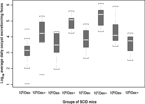
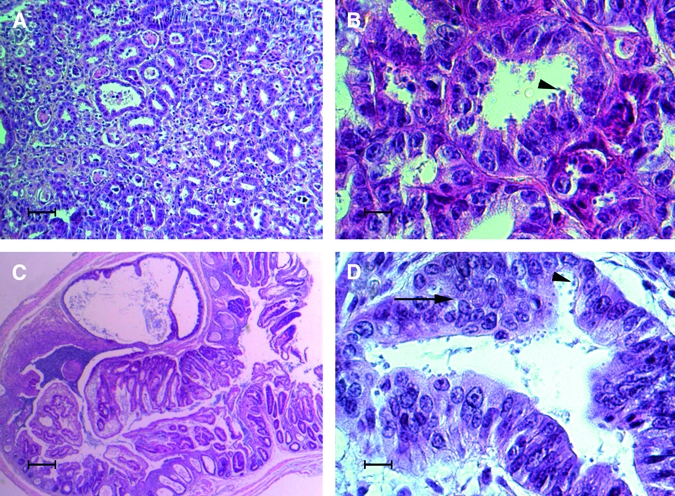
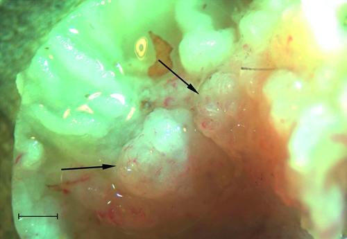
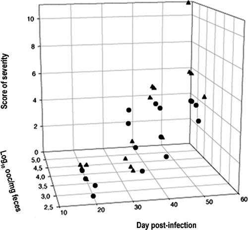
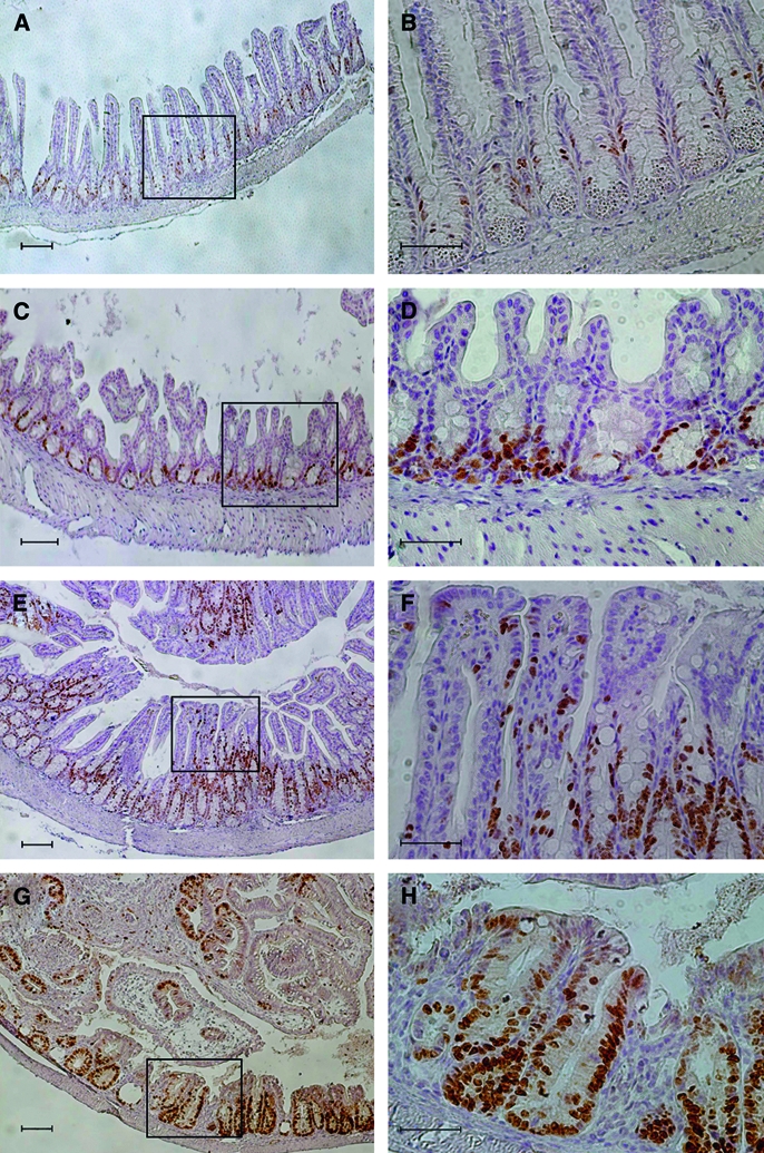
We reported previously that Cryptosporidium parvum was able to induce intestinal tumors in severe combined immunodeficiency (SCID) mice treated with corticoids. To further characterize this Cryptosporidium-induced cell transformation, SCID mice treated with dexamethasone were challenged with C. parvum oocysts, and euthanatized sequentially after infection for histologic examination. Ki-67 was used as a marker of cellular proliferation. Our previous results were confirmed, and it was also found that mice receiving higher inocula (10(6)-10(7)) experienced more severe neoplastic development. Additionally, neoplastic changes were observed not only in the caecum but also in the stomach and duodenum of some animals. Interestingly, SCID mice (6/6) inoculated with 10(5)-10(7) oocysts showed high grade intraepithelial neoplasia or adenomas with high grade dysplasia in the caecum after Day 46 post-infection (PI). Immunohistochemistry for Ki-67 staining indicated the neoplastic process associated to cryptosporidiosis, and evidenced the first immunohistochemical alterations at early stages of the process, even at 3 weeks PI.


Figures

Figure 1.
Figure 1.
Daily Cryptosporidium oocyst excretion (natural logarithm of oocyst/mg feces) in different groups of severe combined immunodeficiency (SCID) mice over the 57 days of the study. Each box represents the median 50% of data, white line is the median. Whiskers represent the extreme values within 1.5 times the box height. Extra lines above and below the whiskers are the outliers.
Figure 2.
Figure 2.
Dex-treated severe combined immunodeficiency (SCID) mice infected with 106 oocysts of C. parvum (euthanasia Day 57 post-infection [PI]). A, Antro-pyloric region showing high-grade intraepithelial neoplasia characterized by an architectural distortion of glands (Bar = 80 μm). B, Antro-pyloric region showing cellular atypias, loss of normal polarity of epithelial cells, and presence of numerous parasites inside the glands (Bar = 15 μm). C, Ileocaecal region showing a projection of a polypoid structure with focal cystic dilation developing inside the intestinal lumen (Bar = 200 μm). D, Areas of high-grade dysplasia (arrow) inside a neoplastic polypoid structure with numerous parasites (arrowhead) (Bar = 15 μm), Hematoxylin & Eosin.
Figure 3.
Figure 3.
Ileocaecal region of a severe combined immunodeficiency (SCID) mouse infected with 105 oocysts of C. parvum and euthanatized after Day 45 PI. Arrows show the presence of adenomatous masses in the intestinal lumen (Bar = 1,000 µm).
Figure 4.
Figure 4.
Correlation between the severity of histologic lesions and the intensity of daily oocyst excretion. Each point represents one mouse. Circles: Dex-untreated mice, Triangles: Dex-treated mice. Note that most animals euthanatized between Days 20 and 35 post-infection [PI] have a score of severity < 2 (see Materials and Methods). All animals euthanatized after Day 35 PI had a severity score ≥ 2 (= neoplastic lesions). There was a significant correlation between severity score and oocyst excretion (r = 0,622, P < 0.001).
Figure 5.
Figure 5.
Expression of Ki-67 in the epithelium of Dex-treated severe combined immunodeficiency (SCID) mice infected with C. parvum. A, Dex-treated uninfected mouse (control) euthanatized at Day 57 post-infection (PI): Ki-67 immunoreactive nuclei are observed at the lower third of the crypts (2–6 immunoreactive cells per glandular section) (Bar = 100 µm). B, Detail of A (delimited area) (Bar = 50 µm). C, Infected mouse euthanatized at Day 20 PI: Ki-67 immunoreactive cells confined to the lower third of the crypts (7–10 immunoreactive cells per glandular section) (Bar = 100 µm). D, Detail of C (delimited area) (Bar = 50 µm). E, Infected mouse euthanatized at Day 35 PI: increased extension of the staining (20–30 immunoreactive cells per glandular section) (Bar = 100 µm). F, Detail of E (delimited area). Proliferating cells are observed in the upper third of the crypts and in the surface epithelium (Bar = 50 µm). G, Infected mouse euthanatized at Day 57 PI: Ki-67 immunoreactive cells are present all over the crypt including the surface epithelium. The architecture of the mucosa is highly altered by the neoplastic process (Bar = 100 µm). H, Detail of G (delimited area) (Bar = 50 µm).

Similar articles

Cited by

References

    1. Sunnotel O, Lowery CJ, Moore JE, Dooley JS, Xiao L, Millar BC, Rooney PJ, Snelling WJ. Cryptosporidium. Lett Appl Microbiol. 2006;43:7–16. - PubMed
    1. Pozio E, Morales MA. The impact of HIV-protease inhibitors on opportunistic parasites. Trends Parasitol. 2005;21:58–63. - PubMed
    1. Okhuysen PC, Chappell CL. Cryptosporidium virulence determinants–are we there yet? Int J Parasitol. 2002;32:517–525. - PubMed
    1. Izquierdo J, Antunez I, Calderon MT, Perez Giraldo C, Munoz Sanz A. Diarrhea caused by Cryptosporidium and colonic neoplasia. Rev Clin Esp. 1988;182:393–394. - PubMed
    1. Sulzyc-Bielicka V, Kuzna-Grygiel W, Kolodziejczyk L, Bielicki D, Kladny J, Stepien-Korzonek M, Telatynska-Smieszek B. Cryptosporidiosis in patients with colorectal cancer. J Parasitol. 2007;93:722–724. - PubMed

Publication types

MeSH terms