

Review
doi: 10.1152/physiol.00038.2009.
Phagocytosis of retinal rod and cone photoreceptors
Affiliations
- PMID: 20134024
- PMCID: PMC2839896
- DOI: 10.1152/physiol.00038.2009
Item in Clipboard
Review
Phagocytosis of retinal rod and cone photoreceptors
Physiology (Bethesda).
2010 Feb.
Abstract
Photoreceptor cells maintain a roughly constant length by continuously generating new outer segments from their base while simultaneously releasing mature outer segments engulfed by the retinal pigment epithelium (RPE). Thus postmitotic RPE cells phagocytose an immense amount of material over a lifetime, disposing of photoreceptor cell waste while retaining useful content. This review focuses on current knowledge of outer segment phagocytosis, discussing the steps involved along with their critical participants as well as how various perturbations in outer segment (OS) disposal can lead to retinopathies.
Figures
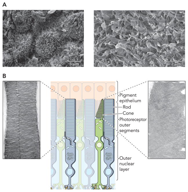
A: high-resolution electron micrograph of RPE cells (left) with a higher resolution image of microvilli on the apical RPE surface (right). Scale bars represent 5 and 2 μm in the low- and high-resolution images. B: graphical representation of rod and cone structures accompanied by EM images of rod and cone outer segments that display the bands and invaginations of these photoreceptors (adapted from Ref. 60).

A: radioautographs of photoreceptor cells showing progression of a radioactive protein band during renewal. Numbered panels display time after injections: 0.5 h (2), 2 days (3), 3 days (4), 4 days (5), 1 wk (6), and 9 days (7). Arrow in panel 7 is pointing to a shed packet of outer segment. B: representation of radioactive band progression during photoreceptor renewal (adopted from Ref. 90).
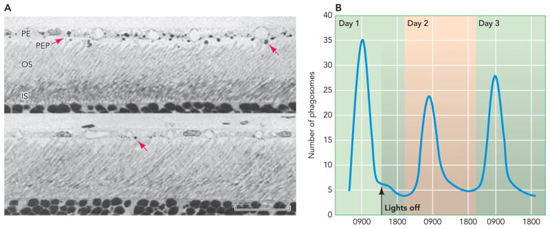
A: light micrographs of rat retinas taken at different times of the day: a. 1.75 h after light onset (a) and 9 h after light onset (b). Arrows in a and b point to phagosomes stained with toluidine blue. B: counts of phagosomes throughout a day and for two days after lights were turned off. Shaded area represents time when lights are off (adapted from Ref. 52). PE, pigmented epithelium; PEP, pigmented epithelial cell processes; OS, outer segment; IS, inner segment.
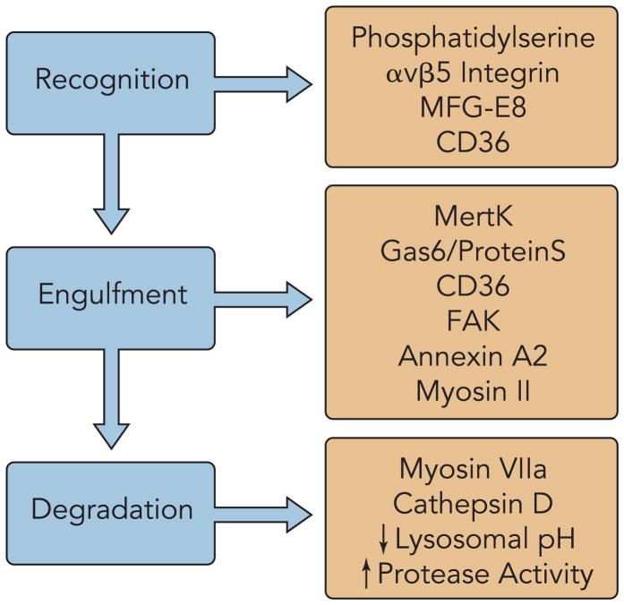
Components are categorized by their involvement in photoreceptor recognition, engulfment, and degradation separated by their involvement in different stages of the renewal process.
References
-
- Anderson DH, Fisher SK. Disc shedding in rodlike and cone-like photoreceptors of tree squirrels. Science. 1975;187:953–955. - PubMed
-
- Anderson DH, Fisher SK, Steinberg RH. Mammalian cones: disc shedding, phagocytosis, renewal. Invest Ophthalmol Vis Sci. 1978;17:117–133. - PubMed
-
- Basinger S, Hoffman R, Matthes M. Photoreceptor shedding is initiated by light in the frog retina. Science. 1976;194:1074–1076. - PubMed
-
- Besharse JC, Hollyfield JG. Turnover of mouse photoreceptor outer segments in constant light and darkness. Invest Ophthalmol Vis Sci. 1979;18:1019–1024. - PubMed
Publication types
MeSH terms
Grants and funding
LinkOut - more resources
Full Text Sources
Other Literature Sources
Medical