

Review
doi: 10.1152/physiol.00042.2009.
Stretch-activated ion channels: what are they?
Affiliations
- PMID: 20134028
- PMCID: PMC2924431
- DOI: 10.1152/physiol.00042.2009
Item in Clipboard
Review
Stretch-activated ion channels: what are they?
Physiology (Bethesda).
2010 Feb.
Abstract
Mechanosensitive ion channels (MSCs) exist in all cells, but mechanosensitivity is a phenotype not a genotype. Specialized mechanoreceptors such as the hair cells of the cochlea require elaborate mechanical impedance matching to couple the channels to the external stress. In contrast, MSCs in nonspecialized cells appear activated by stress in the bilayer local to the channel--within about three lipids. Local mechanical stress can be produced by far-field tension, amphipaths, phase separations, the cytoskeleton, the extracellular matrix, and the adhesion energy between the membrane and a patch pipette. Understanding MSC function requires under standing the stimulus.
Figures
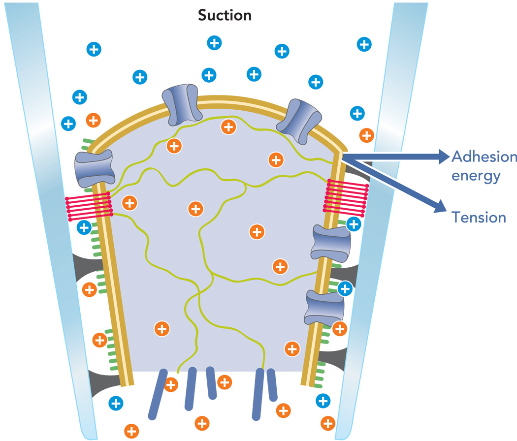
The bilayer is tan, the cytoplasm is light blue with green and dark blue filaments, ion channels are blue tubes crossing the bilayer, the glycocalyx is green, proteins denatured against the glass are black, extracellular cations are in blue, intracellular cations are in orange, and the blue vectors show the force resolution into membrane tension and adhesion energy. Not shown is the upward component of force parallel to the pipette that leads to creep. The postulated lipid seal region where the membrane lipids bind directly to the glass is shown as red bands.
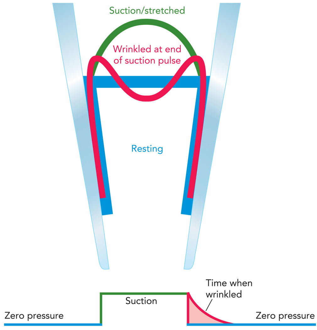
The resting patch (blue trace) is approximately normal to the pipette due to the adhesion energy and has an area of πr2. Applying a step of suction stretches the patch by bringing in new membrane and peeling it off the wall (green trace) creating a spherical cap of area of ~2πrh. Returning the pressure to zero causes the excess membrane area to wrinkle (red trace) until it reanneals to the glass returning to the resting configuration (blue trace). This peeling and reannealing can be seen from changes in patch capacitance (c.f. Figure 3). Thus, although it is wrinkled, the patch has less than resting (baseline) tension.
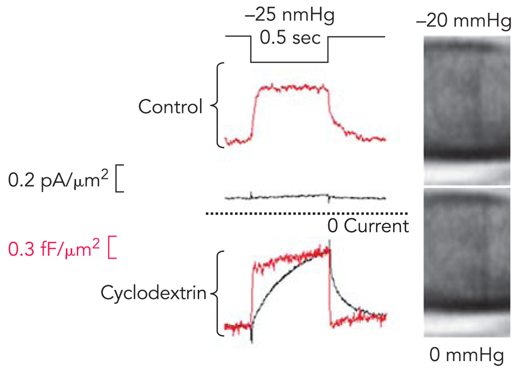
Top traces shows the response of a control cell to a step of suction. The MSC current is small and activates and relaxes slowly (bottom black trace). The patch capacitance, a measure of bilayer stress, increases faster than current, but at a clearly resolvable speed (red trace). Bottom: the response in a patch after treatment of the cells for 2 h with 5mM β methylcyclodextrin. The micrographs at the right of this patch show that patch becomes almost invisible due to the loss of bound proteins. (The images have been digitally enhanced to improve contrast, and the pipette is shown horizontal with the tip to the right). The MSC current is now much larger, although still displaying the activation and deactivation kinetics. The local stress viewed through the patch capacitance (red trace) now changes as fast as the stimulus pulse. The large resting current observed with cyclodextrin is the result of MSC activation by resting tension (Suchyna T, Sachs F, unpublished observations).
References
-
- Andersen OS, Nielsen C, Maer AM, Lundb’k JA, Goulian M, Koeppe RE. Ion channels as tools to monitor lipid bilayer-membrane protein interactions: gramicidin channels as molecular force transducers. Methods Enzymol. 1999;294:208–224. - PubMed
-
- Beech DJ. TRPC1: store-operated channel and more. Pflügers Arch. 2005;451:53–60. - PubMed
-
- Besch SR, Suchyna T, Sachs F. High-speed pressure clamp. Pflügers Arch. 2002;445:161–166. - PubMed
-
- Bett GCL, Sachs F. Whole-cell mechanosensitive currents in rat ventricular myocytes activated by direct stimulation. J Membr Biol. 2000;173:255–263. - PubMed
Publication types
MeSH terms
Substances
Grants and funding
LinkOut - more resources
Full Text Sources
Molecular Biology Databases