

Free-hand thoracic pedicle screws placed by neurosurgery residents: a CT analysis
- PMID: 20135332
- PMCID: PMC2899961
- DOI: 10.1007/s00586-010-1293-1
Free-hand thoracic pedicle screws placed by neurosurgery residents: a CT analysis
Abstract
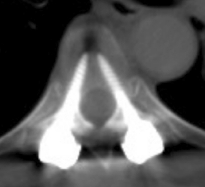
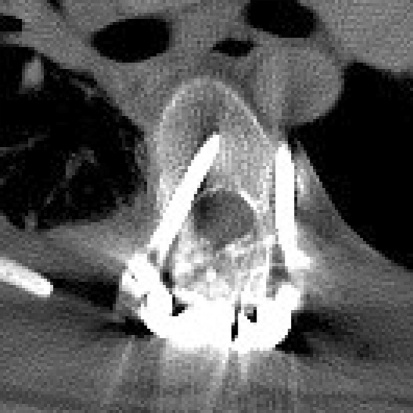
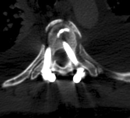
Free-hand thoracic pedicle screw placement is becoming more prevalent within neurosurgery residency training programs. This technique implements anatomic landmarks and tactile palpation without fluoroscopy or navigation to place thoracic pedicle screws. Because this technique is performed by surgeons in training, we wished to analyze the rate at which these screws were properly placed by residents by retrospectively reviewing the accuracy of resident-placed free-hand thoracic pedicle screws using computed tomography imaging. A total of 268 resident-placed thoracic pedicle screws was analyzed using axial computed tomography by an independent attending neuroradiologist. Eighty-five percent of the screws were completely within the pedicle and that 15% of the screws violated the pedicle cortex. The majority of the breaches were lateral breaches between 2 and 4 mm (46%). There was no clinical evidence of neurovascular injury or injury to the esophagus. There were no re-operations for screw replacement. We concluded that under appropriate supervision, neurosurgery residents can safely place free-hand thoracic pedicle screws with an acceptable breach rate.
Figures
References
-
- Liljenqvist U, Hackenberg L, Link T, Halm H. Pullout strength of pedicle screws versus pedicle and laminar hooks in the thoracic spine. Acta Orthop Belg. 2001;67:157–163. - PubMed
-
- Vaccaro AR, Rizzolo SJ, Allardyce TJ, et al. Placement of pedicle screws in the thoracic spine. Part I: morphometric analysis of the thoracic vertebrae. J Bone Joint Surg Am. 1995;77:1193–1199. - PubMed
