

Glycosaminoglycans modulate RANKL-induced osteoclastogenesis
- PMID: 20135643
- PMCID: PMC3095103
- DOI: 10.1002/jcb.22506
Glycosaminoglycans modulate RANKL-induced osteoclastogenesis
Abstract
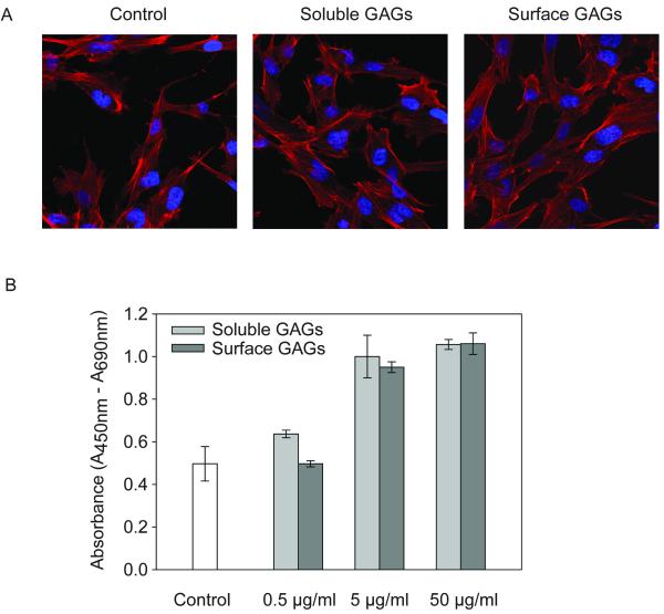
Skeletal integrity is tightly regulated by the activity of osteoblasts and osteoclasts that are both under the control of extracellular glycosaminoglycans (GAGs) through their interactions with endogenous growth factors and differentiation-promoting ligands. Receptor activator of NF-kappa-B ligand (RANKL), which is a tumor necrosis factor (TNF)-related protein that is critical for osteoclast formation, is produced by osteoblasts and further modulated by certain types of GAGs. Using unfractionated osteoblast-derived GAGs that reflect the complex tissue microenvironment within which osteoclasts reside, we demonstrate that these GAGs block the osteoclastogenic activity of RANKL. Furthermore, RANKL significantly reduces extracellular signal-regulated protein kinase (ERK) activity, a putative suppressor of osteoclastogenesis, but osteoblast-derived GAGs eliminate the inhibitory effects of RANKL on ERK activity. Notably, while imposing an anti-osteoclastic effect, these GAGs also enhanced the proliferation of osteoblasts. Thus, the osteoblast microenvironment is a potent source of GAGs that promote bone anabolic activities. The anti-osteoclastogenic and osteoblast-related mitogenic activities of these GAGs together may provide a key starting point for the development of selective sugar-based therapeutic compounds for the treatment of osteopenic disorders.
Figures


Similar articles
-
Glycosaminoglycans inhibit the adherence and the spreading of osteoclasts and their precursors: role in osteoclastogenesis and bone resorption.Eur J Cell Biol. 2011 Jan;90(1):49-57. doi: 10.1016/j.ejcb.2010.08.001. Epub 2010 Oct 20. Eur J Cell Biol. 2011. PMID: 20970218
-
Heparin inhibits osteoclastic differentiation and function.J Cell Biochem. 2008 Apr 15;103(6):1707-17. doi: 10.1002/jcb.21559. J Cell Biochem. 2008. PMID: 18231993
-
Caffeic acid 3,4-dihydroxy-phenethyl ester suppresses receptor activator of NF-κB ligand–induced osteoclastogenesis and prevents ovariectomy-induced bone loss through inhibition of mitogen-activated protein kinase/activator protein 1 and Ca2+–nuclear factor of activated T-cells cytoplasmic 1 signaling pathways.J Bone Miner Res. 2012 Jun;27(6):1298-1308. doi: 10.1002/jbmr.1576. J Bone Miner Res. 2012. PMID: 22337253
-
Osteoclast differentiation by RANKL and OPG signaling pathways.J Bone Miner Metab. 2021 Jan;39(1):19-26. doi: 10.1007/s00774-020-01162-6. Epub 2020 Oct 20. J Bone Miner Metab. 2021. PMID: 33079279 Review.
-
A new member of tumor necrosis factor ligand family, ODF/OPGL/TRANCE/RANKL, regulates osteoclast differentiation and function.Biochem Biophys Res Commun. 1999 Mar 24;256(3):449-55. doi: 10.1006/bbrc.1999.0252. Biochem Biophys Res Commun. 1999. PMID: 10080918 Review.
Cited by
-
Regenerative potential of glycosaminoglycans for skin and bone.J Mol Med (Berl). 2012 Jun;90(6):625-35. doi: 10.1007/s00109-011-0843-2. Epub 2011 Dec 21. J Mol Med (Berl). 2012. PMID: 22187113 Review.
-
Glycosaminoglycan mimetic associated to human mesenchymal stem cell-based scaffolds inhibit ectopic bone formation, but induce angiogenesis in vivo.Tissue Eng Part A. 2013 Jul;19(13-14):1641-53. doi: 10.1089/ten.TEA.2012.0377. Tissue Eng Part A. 2013. PMID: 23521005 Free PMC article.
-
Proteoglycans and osteolysis.Methods Mol Biol. 2012;836:323-37. doi: 10.1007/978-1-61779-498-8_21. Methods Mol Biol. 2012. PMID: 22252644 Free PMC article.
-
Osteoblast-released Matrix Vesicles, Regulation of Activity and Composition by Sulfated and Non-sulfated Glycosaminoglycans.Mol Cell Proteomics. 2016 Feb;15(2):558-72. doi: 10.1074/mcp.M115.049718. Epub 2015 Nov 23. Mol Cell Proteomics. 2016. PMID: 26598647 Free PMC article.
-
Nanoparticulate mineralized collagen glycosaminoglycan materials directly and indirectly inhibit osteoclastogenesis and osteoclast activation.J Tissue Eng Regen Med. 2019 May;13(5):823-834. doi: 10.1002/term.2834. Epub 2019 Apr 15. J Tissue Eng Regen Med. 2019. PMID: 30803152 Free PMC article.
References
-
- Ariyoshi W, Takahashi T, Kanno T, Ichimiya H, Shinmyouzu K, Takano H, Koseki T, Nishihara T. Heparin inhibits osteoclastic differentiation and function. J Cell Biochem. 2008;103:1707–17. - PubMed
-
- Ariyoshi W, Takahashi T, Kanno T, Ichimiya H, Takano H, Koseki T, Nishihara T. Mechanisms involved in enhancement of osteoclast formation and function by low molecular weight hyaluronic acid. J Biol Chem. 2005;280:18967–72. - PubMed
-
- Braunsteiner H, Thumb N. Mast cells, basophils and heparin liberation. Bibl Haematol. 1963;15:9–15. - PubMed
Publication types
MeSH terms
Substances
Grants and funding
LinkOut - more resources
Full Text Sources
Other Literature Sources
Miscellaneous
