. 2010 Apr;28(4):799-809.
doi: 10.1002/stem.316.

Hypoxia influences the vascular expansion and differentiation of embryonic stem cell cultures through the temporal expression of vascular endothelial growth factor receptors in an ARNT-dependent manner

Affiliations

Hypoxia influences the vascular expansion and differentiation of embryonic stem cell cultures through the temporal expression of vascular endothelial growth factor receptors in an ARNT-dependent manner

Yu Han et al. Stem Cells. 2010 Apr.

Abstract

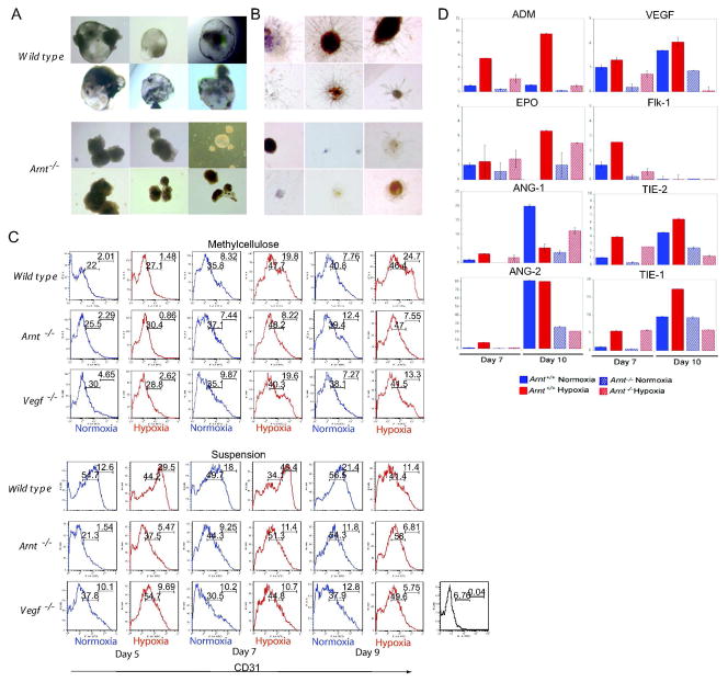
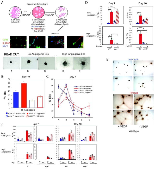
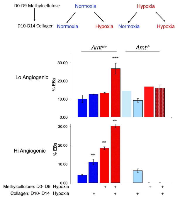
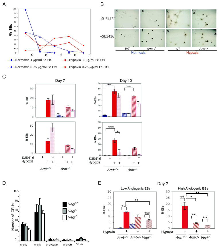
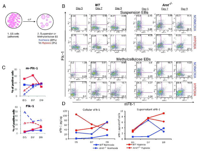
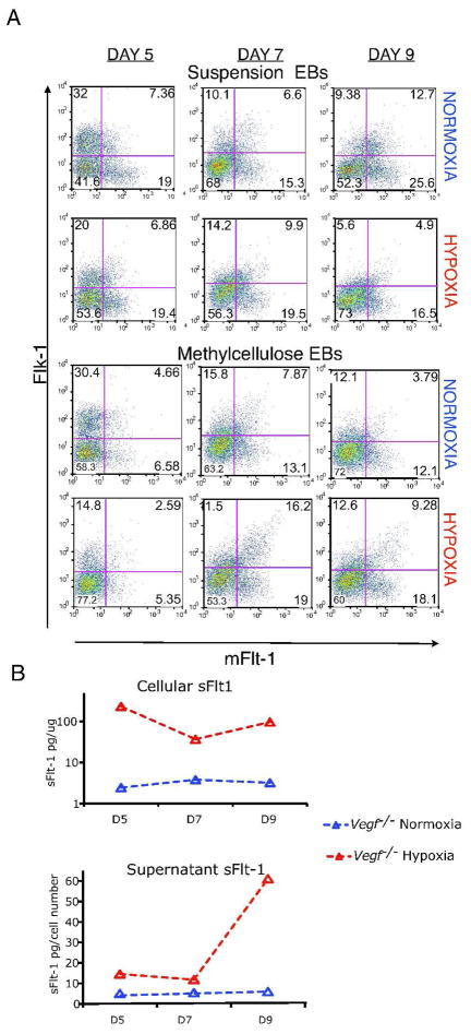
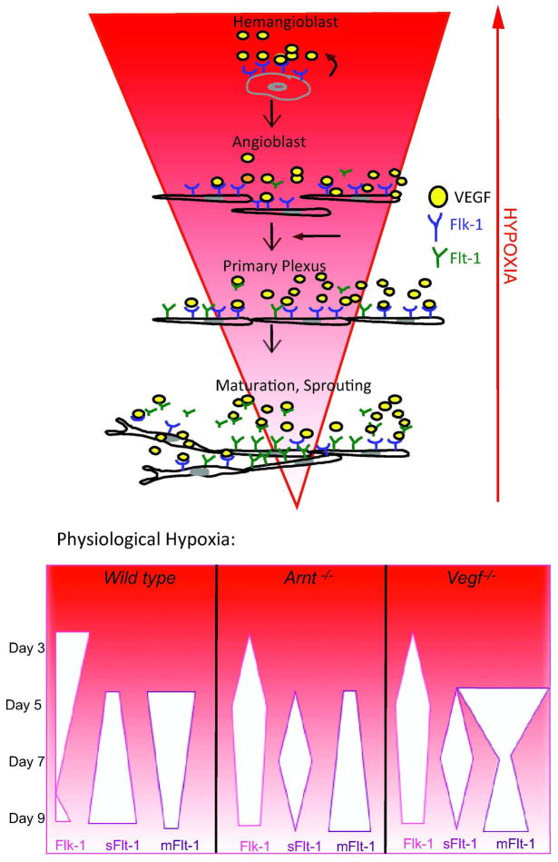
Adaptive responses to low oxygen (O(2)) tension (hypoxia) are mediated by the heterodimeric transcription factor hypoxia inducible factor (HIF). When stabilized by hypoxia, bHLH-PAS alpha- and beta- (HIF-1beta or ARNT) HIF complex regulate the expression of multiple genes, including vascular endothelial growth factor (VEGF). To investigate the mechanism(s) through which hypoxia contributes to blood vessel development, we used embryonic stem cell (ESC) differentiation cultures that develop into embryoid bodies (EBs) mimicking early embryonic development. Significantly, low O(2) levels promote vascular development and maturation in wild-type (WT) ESC cultures measured by an increase in the numbers of CD31(+) endothelial cells (ECs) and sprouting angiogenic EBs, but refractory in Arnt(-/-) and Vegf(-/-) ESC cultures. Thus, we propose that hypoxia promotes the production of ECs and contributes to the development and maturation of vessels. Our findings further demonstrate that hypoxia alters the temporal expression of VEGF receptors Flk-1 (VEGFR-2) and the membrane and soluble forms of the antagonistic receptor Flt-1 (VEGFR-1). Moreover, these receptors are distinctly expressed in differentiating Arnt(-/-) and Vegf(-/-) EBs. These results support existing models in which VEGF signaling is tightly regulated during specific biologic events, but also provide important novel evidence that, in response to physiologic hypoxia, HIF mediates a distinct stoichiometric pattern of VEGF receptors throughout EB differentiation analogous to the formation of vascular networks during embryogenesis.


Figures

Figure 1
Figure 1
Loss of Arnt disrupts proper vascular differentiation in ES differentiation cultures. (A,B) Arnt−/− ESCs stained for CD31 demonstrate a failure to generate vascular cystic embryoid bodies in 20 day EBs grown in suspension (A) or sprouting EBs differentiated in Type I collagen (B) compared to WT controls. (C) Flow cytometry was used to assess CD31 levels from cells obtained by dissociating EBs from WT, Arnt−/−, or Vegf−/− ESCs differentiated in methylcellulose or suspension under normoxia or hypoxia (3% O2). (D) Transcript levels of angiogenic genes from cDNA isolated from WT or Arnt−/− Day 7 and 10 EBs were determined by real-time PCR, normalized against 18S, and expressed as mean±SEM relative to the mean of D7 WT normoxia sample.
Figure 2
Figure 2
The presence of ARNT in response to hypoxia influences angiogenic growth in differentiating ESC cultures independent of exogenous VEGF. (A) Experimental model used to differentiate EBs and quantify their angiogenic potential. Representative fields of vessels sprouts immunostained to reflect vessels containing CD31+ EC and α-SMA+ vascular smooth muscle cells. At least 50 individual EBs replated in 3D Type I Collagen are classified as Low or High angiogenic EBs. Experiments were performed in triplicate and data represented as percent mean values ±SEM, *p< .05, **p< .005, *p< .001. (B–E) Quantitative analysis of angiogenesis from WT or Arnt−/− ES cells differentiated under normoxia or hypoxia (3% O2) and replated in Type I collagen. (B) Percent of high angiogenic EBs of cultures differentiated for 10 Days in the presence of 25 ng/ml VEGF and 100 ng/ml bFGF (C) Results from similar EB cultures differentiated for 7 days with growth factors reduced to 5 ng/ml VEGF and 25 ng/ml bFGF. (D, E) Results from EB differentiated for 7 or 10 days in the absence of exogenous VEGF. Representative micrographs of 10 day WT cultures grown under normoxia (a,b,c,d) or hypoxia (e,f,g,h) with (a,b,e,f) or without (c,d,g,h) exogenous VEGF and replated in Type I collagen to assess angiogenic potential. (F) WT or Arnt−/− ES cells differentiated in methylcellulose with 25, 5, or 0 ng/ml VEGF for 7 or 10 days and then replated in Type I collagen and individual EBs were scored for displaying low or high angiogenic outgrowth.
Figure 3
Figure 3
Hypoxia promotes vascular differentiation and angiogenic sprouting in an ARNT-dependent manner. (A) Experimental model outlining the normoxic or hypoxic (3% O2) conditions used for the differentiation of EBs in methylcellulose (in the absence of exogenous VEGF) and during vascular outgrowth in Type I collagen. (B) Individual EBs were scored for angiogenic outgrowth for each condition. Data represent mean±SEM from triplicate experiments, **p< .01, ***p< .001.
Figure 4
Figure 4
Hypoxia promotes vascular differentiation independent of VEGF. (A,B,C) Inhibiting VEGF is not sufficient to block hypoxia’s angiogenic effects. Inhibitors were added during the differentiation of EBs for 7 or 10 days in methylcellulose EBs exposed to normoxic or hypoxic (3% O2) conditions and at least 50 EBs per treatment were then replated in Type I Collagen and EBs were individually scored for angiogenic outgrowth. (A) Fc-Flt1 was used at 1 and 0.25 μg/ml, and results are represented as percent of each EB angiogenic type from total numbers of EBs per treatment. (B,C) WT or Arnt−/− EBs were differentiated under normoxic or hypoxic conditions with and without with 50 μM SU5416 and replated in Type I collagen. (B) Micrographs of representative cultures demonstrate that WT EBs preserve some angiogenic EBs. (C) Quantification of Low and High angiogenic EBs in response to SU5416 treatment. Data represent means from triplicate samples ± SEM. (D,E) Vegf−/− ESCs contain all hematopoietic colony forming potential but are angiogenic deficient. (D) Hematopoietic progenitor activity of day 9 Vegf−/−, Vegf+/−, and WT EB-derived cells. 1500 cells were replated in triplicate and individual colonies were scored 6–7 days after replating. Error bars represent SEM. (E) Quantitative analysis of angiogenesis from WT, Arnt−/−, or Vegf−/− ES cells differentiated for 7 days under normoxia or hypoxia (3% O2) in the absence of exogenous VEGF. Over 50 EBs replated in Type I collagen in triplicate and individual colonies were scored for angiogenic sprouting.
Figure 5
Figure 5
Hypoxia regulates the temporal expression of VEGF receptors. (A) EBs were differentiated in suspension or methylcellulose cultures in the absence of exogenous VEGF. Cultures were exposed to normoxia or hypoxia (3% O2) and analyzed for the surface expression of Flk-1 or mFlt-1 by flow cytometry (B,C) and secreted Flt-1 by ELISA (D). (C) In WT cultures, hypoxia (solid red squares) alters the expression of each receptor compared to normoxic conditions (solid blue squares) and are further dysregulated in Arnt−/− EB cultures. (D) Although distinct from each other, the concentration of sFlt-1 from protein cell lysates isolated from methylcellulose EB cultures and conditioned media from suspension EB cultures is increased by hypoxic treatment in both WT and Arnt−/− EB cultures.
Figure 6
Figure 6
(A) Vegf−/− ES were differentiated in suspension or methylcellulose in the absence of exogenous VEGF. Cultures were exposed to normoxia or hypoxia (3% O2) and dissociated cells were analyzed for the surface expression of Flk-1 or mFlt-1 by flow cytometry. (B) Concentrations of secreted Flt-1 from supernatant or protein lysates of Vegf−/− EB cultures were analyzed by ELISA.
Figure 7
Figure 7
Model illustrating that hypoxia influences the expression of VEGF receptors either promoting (Flk-1) or inhibiting (sFlt-1) vessel sprouting, thereby controlling the formation of vascular networks. Angiogenic defects in absence of Arnt or Vegf may result in part from the altered expression of these receptors.

Similar articles

Cited by

References

    1. Risau W. Mechanisms of angiogenesis. Nature. 1997;386:671–674. - PubMed
    1. Semenza GL. Regulation of oxygen homeostasis by hypoxia-inducible factor 1. Physiology (Bethesda) 2009;24:97–106. - PubMed
    1. Simon MC, Liu L, Barnhart BC, et al. Hypoxia-induced signaling in the cardiovascular system. Annu Rev Physiol. 2008;70:51–71. - PMC - PubMed
    1. Ramirez-Bergeron DL, Runge A, Dahl KD, et al. Hypoxia affects mesoderm and enhances hemangioblast specification during early development. Development. 2004;131:4623–4634. - PubMed
    1. Ramirez-Bergeron DL, Runge A, Adelman DM, et al. HIF-dependent hematopoietic factors regulate the development of the embryonic vasculature. Dev Cell. 2006;11:81–92. - PMC - PubMed

Publication types

MeSH terms

Substances