. 2010 Mar 15;50(6):850-9.
doi: 10.1086/650581.

Delayed clearance of viral load and marked cytokine activation in severe cases of pandemic H1N1 2009 influenza virus infection

Affiliations

Delayed clearance of viral load and marked cytokine activation in severe cases of pandemic H1N1 2009 influenza virus infection

Kelvin K W To et al. Clin Infect Dis. .

Abstract

Background: Infections caused by the pandemic H1N1 2009 influenza virus range from mild upper respiratory tract syndromes to fatal diseases. However, studies comparing virological and immunological profile of different clinical severity are lacking.

Methods: We conducted a retrospective cohort study of 74 patients with pandemic H1N1 infection, including 23 patients who either developed acute respiratory distress syndrome (ARDS) or died (ARDS-death group), 14 patients with desaturation requiring oxygen supplementation and who survived without ARDS (survived-without-ARDS group), and 37 patients with mild disease without desaturation (mild-disease group). We compared their pattern of clinical disease, viral load, and immunological profile.

Results: Patients with severe disease were older, more likely to be obese or having underlying diseases, and had lower respiratory tract symptoms, especially dyspnea at presentation. The ARDS-death group had a slower decline in nasopharyngeal viral loads, had higher plasma levels of proinflammatory cytokines and chemokines, and were more likely to have bacterial coinfections (30.4%), myocarditis (21.7%), or viremia (13.0%) than patients in the survived-without-ARDS or the mild-disease groups. Reactive hemophagocytosis, thrombotic phenomena, lymphoid atrophy, diffuse alveolar damage, and multiorgan dysfunction similar to fatal avian influenza A H5N1 infection were found at postmortem examinations.

Conclusions: The slower control of viral load and immunodysregulation in severe cases mandate the search for more effective antiviral and immunomodulatory regimens to stop the excessive cytokine activation resulting in ARDS and death.


Figures
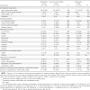
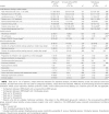
Table 1
Table 1
Demographic Characteristics and Underlying Comorbidities
Table 2
Table 2
Investigations, Treatments, Complications, and Clinical Outcomes
Table 3
Table 3
Initial Plasma Cytokine or Chemokine Levels and Viral Load in Respiratory Specimens
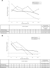
Figure 1
Figure 1
Nasopharyngeal viral load in patients with different clinical severity, according to number of days after symptom onset (A) and to number of days after initiation of oseltamivir treatment (B). Horizontal dotted line indicates detection limit of reverse-transcription polymerase chain reaction. ARDS, acute respiratory distress syndrome.
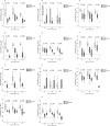
Figure 2
Figure 2
Plasma cytokine or chemokine level, stratified by days after symptom onset. Median, quartiles, and range are shown. The Jonckheere-Terpstra trend test was used to compare cytokine level with disease severity. Horizontal dotted line detection limit of the assay. A Granulocyte colony-stimulating factor. B Interferon (IFN)– lpha;2. C Interleukin (IL)–1 lpha;. D IL-6. E IL-8. F IL-10. G IL-15. H IL-17. I IFN- amma;–induced protein 10. J Monocyte chemoattractant protein–1. K Tumor necrosis factor-α.
Figure 3
Figure 3
Histopathological changes of pulmonary and extrapulmonary tissue (hematoxylin-eosin stained) of the deceased patients suffering from pandemic influenza A H1N1 2009. A Lung parenchyma showing acute exudative phase of diffuse alveolar damage on day 7 after symptom onset in a 42-year-old previously healthy man with interstitial cellular infiltrate and proteinaceous exudates in alveoli and hyaline membrane formation (original magnification, imes;100). B Lung parenchyma showing chronic fibroproliferative phase of diffuse alveolar damage with marked thickening of the alveolar septa and alveolar space being replaced by fibrogranulation tissues on day 28 of symptom onset in a 37-year-old previously healthy woman (original magnification, imes;200). C Thrombosis of branches of pulmonary artery in the same patient in panel B who died despite extracorporeal membrane oxygenation (original magnification, imes;200). D Lymphoid atrophy with no recognizable germinal centre in the spleen of the same patient in panels B and C. The periarteriolar lymphoid sheath (white pulp), which was markedly reduced in size (original magnification, imes;400). E Myocardium showing a microscopic focus of lymphoid aggregate accompanied by myofibril necrosis in a 58-year-old man presenting with elevated creatine kinase isoenzyme CK-MB and echocardiographic evidence of diastolic dysfunction (original magnification, imes;200). F Mediastinal lymph node showing reactive hemophagocytosis in the same patient in panel E (original magnification, imes;400).

Similar articles

Cited by

References

    1. Wong SS, Yuen KY. Avian influenza virus infections in humans. Chest. 2006;129:156–168. - PMC - PubMed
    1. Chowell G, Bertozzi SM, Colchero MA, et al. Severe respiratory disease concurrent with the circulation of H1N1 influenza. N Engl J Med. 2009;361:674–679. - PubMed
    1. Trifonov V, Khiabanian H, Rabadan R. Geographic dependence, surveillance, and origins of the 2009 influenza A (H1N1) virus. N Engl J Med. 2009;361:115–119. - PubMed
    1. Munster VJ, de Wit E, van den Brand JM, et al. Pathogenesis and transmission of swine-origin 2009 A(H1N1) influenza virus in ferrets. Science. 2009;325:481–483. - PMC - PubMed
    1. Maines TR, Jayaraman A, Belser JA, et al. Transmission and pathogenesis of swine-origin 2009 A(H1N1) influenza viruses in ferrets and mice. Science. 2009;325:484–487. - PMC - PubMed

Publication types