. 2010 Apr;216(4):489-95.
doi: 10.1111/j.1469-7580.2009.01203.x. Epub 2010 Jan 28.

Somatotopic organization of lumbar muscle-innervating neurons in the ventral horn of the rat spinal cord

Affiliations

Somatotopic organization of lumbar muscle-innervating neurons in the ventral horn of the rat spinal cord

Yuzuru Takahashi et al. J Anat. 2010 Apr.

Abstract

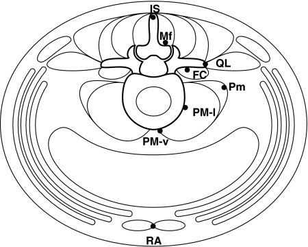
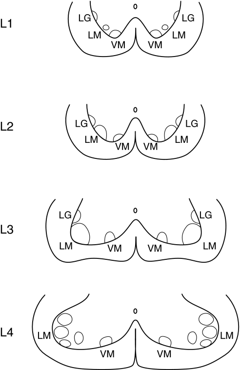
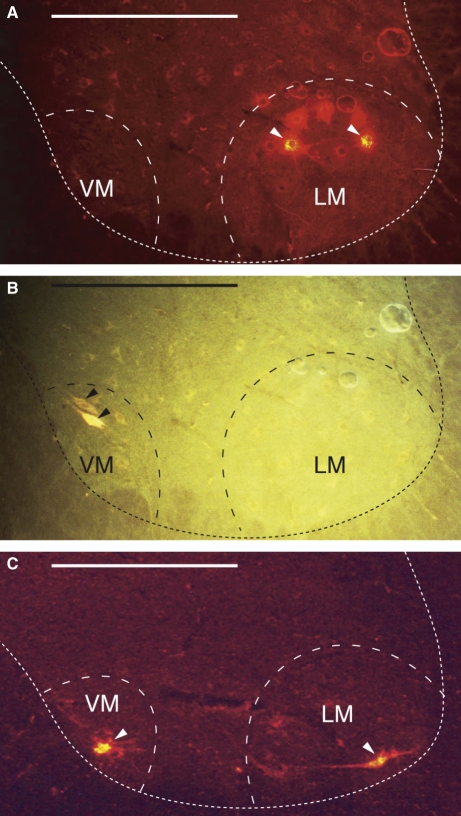
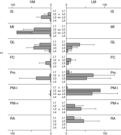
The ventral horn of the rat spinal cord was investigated with respect to the somatotopic organization of the motor neurons that innervate the lumbar muscles. Neurotracer 1,1'-dioctadecyl-3,3,3',3'-tetramethylindocarbocyanine perchlorate (DiI) was applied to specific sites in lumbar muscles. Spinal cord segments at L1 through L4 levels were cut into 40-mum serial transverse sections. Labeled neurons were located in the ventromedial nucleus (VM) and lateromedial nucleus (LM) nuclei of Rexed's lamina IX. Motor neurons innervating the m. interspinales lumborum and m. multifidus were without exception present in the VM, whereas all motor neurons innervating the m. rectus abdominis were present in the LM. Forty percent of motor neurons innervating the m. quadratus lumborum were present in the VM and the other 60% were in the LM. Although most of the motor neurons innervating the m. psoas major were present in the LM, a few labeled neurons existed in the VM. These results suggest that the border zone demarcating the areas of innervation of the dorsal and ventral rami of spinal nerves crosses the m. quadratus lumborum.


Figures

Fig. 1
Fig. 1
Illustration of the sites to which DiI was applied (closed circles). All the sites are located in the transverse plane through the L5 vertebra and L5–L6 intervertebral disc. IS, m. interspinales lumborum at the spinous process of the L5 vertebra; Mf, m. multifidus at the lamina of the L5 vertebra; QL, m. quadratus lumborum at the transverse process of the L5 vertebra; FC, m. flexor caudae at the dorsolateral surface of the L5–L6 intervertebral disc; Pm, m. psoas minor at the L5–L6 intervertebral disc level; PM-l, m. psoas major at the lateral surface of the L5–L6 intervertebral disc; PM-v, m. psoas major at the ventromedian surface of the L5–L6 intervertebral disc; RA, m. rectus abdominis at the pubic synthesis.
Fig. 2
Fig. 2
Three nuclei of Rexed’s lamina IX in the ventral horn of the rat spinal cord L1 through L4 (Molander et al. 1984; Paxinos & Watson, 1998). VM, ventromedial nucleus; LM, lateromedial nucleus; LG, lateral group. Modifications (only the ventral horn is shown) from the original drawings were made by the present authors.
Fig. 3
Fig. 3
Photographic presentation of fluorescent motor neurons in transverse sections of the spinal cord. (A) and (B) are the same section from the spinal cord segment L3 from a rat in which DiI and FG were applied to the m. psoas major (PM-l) and to the m. multifidus (Mf), respectively. (A) Viewed with a G1-A filter. Two DiI-labeled neurons (arrowheads) are identified in the lateromedial nucleus (LM). (B) Viewed with a UV filter. One FG-labeled neuron (arrowheads) is identified in the ventromedial nucleus (VM). (C) A section of the spinal cord segment L3 from a rat in which DiI was applied to the m. quadratus lumborum (QL). DiI-labeled neurons (arrowheads) are present in both the VM and the LM nuclei. The dotted line indicates the contour of the ventral horn. Horizontal bar = 500 μm.
Fig. 4
Fig. 4
Number of labeled neurons in the spinal cord segments L1–L4 and DRGs L1–L4. Data are presented as the mean ± SD. *P < 0.05, **P < 0.01. IS, m. interspinales lumborum at the spinous process of the L5 vertebra; Mf, m. multifidus at the lamina of the L5 vertebra; QL, m. quadratus lumborum at the transverse process of the L5 vertebra; FC, m. flexor caudae at the dorsolateral surface of the L5–L6 intervertebral disc; Pm, m. psoas minor at the L5–L6 intervertebral disc level; PM-l, m. psoas major at the lateral surface of the L5–L6 intervertebral disc; PM-v, m. psoas major at the ventromedial surface of the L5–L6 intervertebral disc; RA, m. rectus abdominis at the pubic synthesis.
Fig. 5
Fig. 5
Number of labeled motor neurons in the ventromedial (VM) and lateromedial (LM) nuclei of lamina IX in the ventral horn of L1–L4 spinal segments. Data are presented as mean ± SD. *P < 0.05, **P < 0.01. IS, m. interspinales lumborum at the spinous process of the L5 vertebra; Mf, m. multifidus at the lamina of the L5 vertebra; QL, m. quadratus lumborum at the transverse process of the L5 vertebra; FC, m. flexor caudae at the dorsolateral surface of the L5–L6 intervertebral disc; Pm, m. psoas minor at the L5–L6 intervertebral disc level; PM-l, m. psoas major at the lateral surface of the L5–L6 intervertebral disc; PM-v, m. psoas major at the ventromedian surface of the L5–L6 intervertebral disc; RA, m. rectus abdominis at the pubic synthesis. Note that application of neurotracer to the QL, FC, Pm, and PM sites produced labeled motor neurons in both the VM or LM nuclei.
Fig. 6
Fig. 6
Illustration of how the dorsoventral axis of the lumbar musculoskeletal tissues is reproduced in the dorsal and ventral horn of the spinal cord. Note that the directions are reversed in the dorsal and ventral horns.

Similar articles

Cited by

References

    1. Agalliu D, Takada S, Agalliu I, et al. Motor neurons with axial muscle projections specified by Wnt4/5 signaling. Neuron. 2009;61:708–720. - PMC - PubMed
    1. Bogduk N. The innervation of the lumbar spine. Spine. 1983;8:286–293. - PubMed
    1. Bogduk N. Nerves of the lumbar spine. In: Bogduk N, editor. Clinical Anatomy of the Lumbar Spine and Sacrum. New York: Churchill Livingstone; 1997. p. 143.
    1. Bogduk N, Tynan W, Wilson AS. The nerve supply to the human lumbar intervertebral discs. J Anat. 1981;132:39–56. - PMC - PubMed
    1. Brown PB, Fuchs JL. Somatotopic representation of hindlimb skin in cat dorsal horn. J Neurophysiol. 1975;38:1–9. - PubMed

LinkOut - more resources