. 2010 Jun;160(3):604-14.
doi: 10.1111/j.1476-5381.2009.00625.x. Epub 2010 Feb 5.

GPR55 ligands promote receptor coupling to multiple signalling pathways

Affiliations

GPR55 ligands promote receptor coupling to multiple signalling pathways

Christopher M Henstridge et al. Br J Pharmacol. 2010 Jun.

Abstract

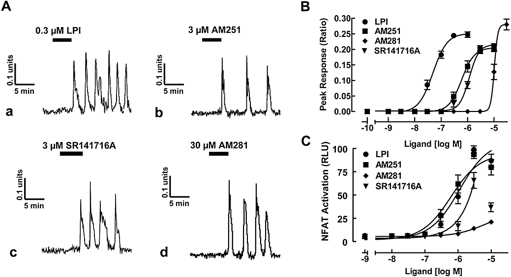
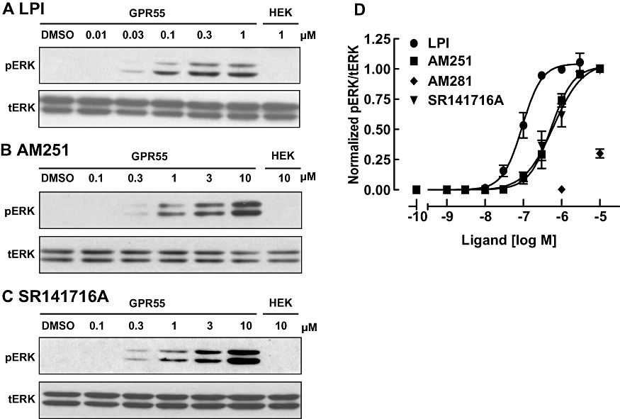
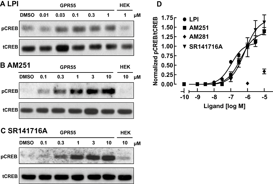
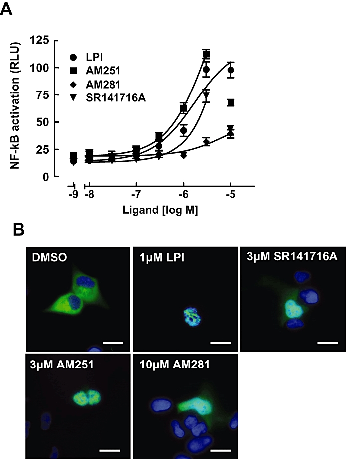
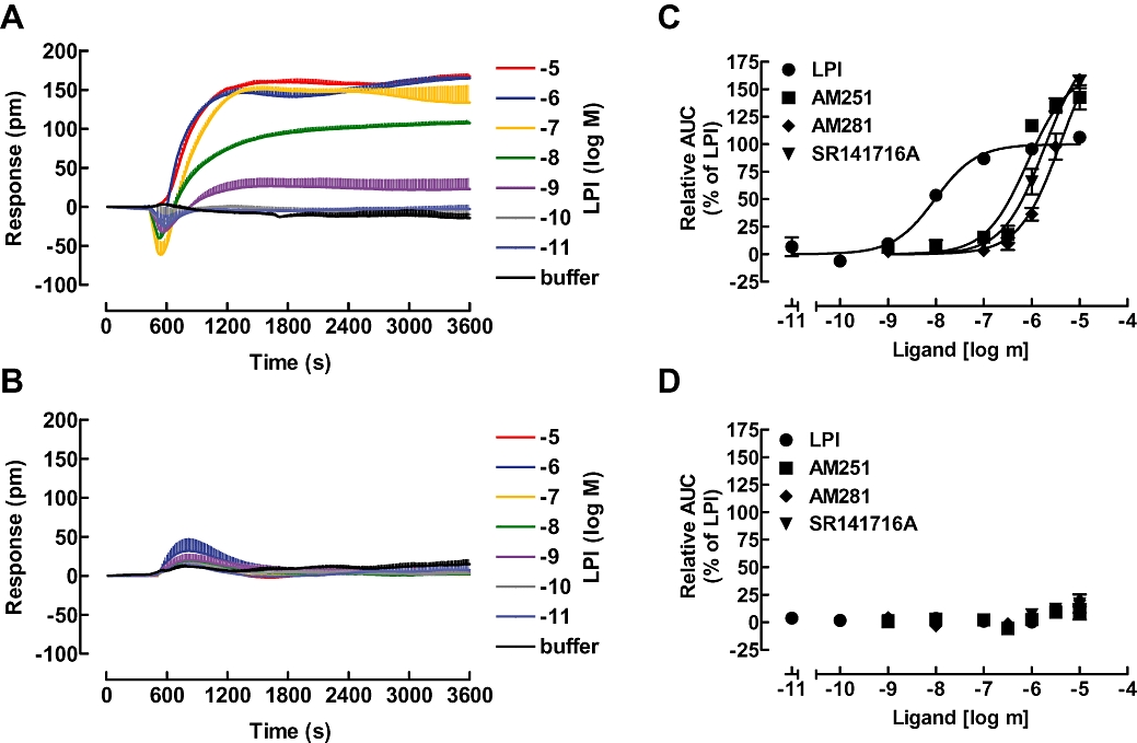
Background and purpose: Although GPR55 is potently activated by the endogenous lysophospholipid, L-alpha-lysophosphatidylinositol (LPI), it is also thought to be sensitive to a number of cannabinoid ligands, including the prototypic CB1 receptor antagonists AM251 and SR141716A (Rimonabant). In this study we have used a range of functional assays to compare the pharmacological activity of selected cannabinoid ligands, AM251, AM281 and SR141716A with LPI in a HEK293 cell line engineered to stably express recombinant, human GPR55.

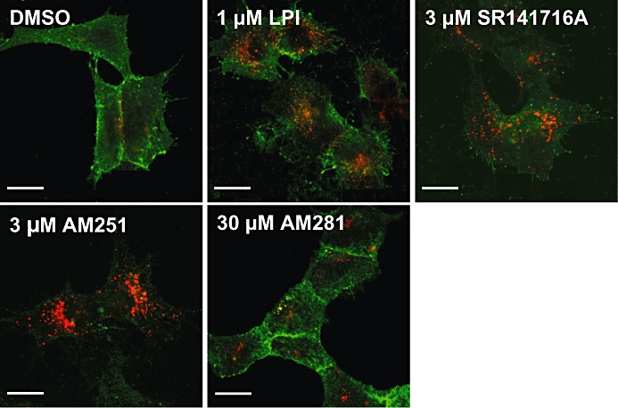
Experimental approach: We evaluated Ca(2+) signalling, stimulation of extracellular signal regulated kinase (ERK1/2) mitogen activated kinase MAP-kinases, induction of transcriptional regulators that are downstream of GPR55, including nuclear factor of activated T cells (NFAT), nuclear factor-kappaB (NF-kappaB) and cAMP response element binding protein (CREB), as well as receptor endocytosis. In addition, we assessed the suitability of a novel, label-free assay for GPR55 ligands that involves optical measurement of dynamic mass redistribution following receptor activation.

Key results: GPR55 linked to a range of downstream signalling events and that the activity of GPR55 ligands was influenced by the functional assay employed, with differences in potency and efficacy observed.

Conclusions and implications: Our data help to resolve some of the issues surrounding the pharmacology of cannabinoid ligands at GPR55 and highlight some differences in effector coupling associated with distinct GPR55 ligands.


Figures

Figure 1
Figure 1
GPR55 ligands promote oscillatory Ca2+ transients and NFAT-activation in GPR55-HEK293 cells. (A) Representative Ca2+ transients in GPR55-HEK293 cells. (a) LPI (0.3 µM), (b) AM251 (3 µM), (c) SR141716A (3 µM) and (d) AM281 (30 µM) induce a sustained, oscillatory Ca2+ response in GPR55-HEK293 cells. Traces represent changes in fura-2 fluorescence ratio for individual cells. (B) Concentration–response curves for GPR55 ligands. LPI, AM251, SR141716A and AM281 all induced Ca2+ transients. Data are mean peak responses ± SEM of 40 cells derived from four independent experiments. (C) NFAT transcription factor activation in GPR55-expressing cells. GPR55-HEK293 cells were transfected with 200 ng of NFAT-luciferase-reporter plasmid and 24 h post transfection, cells were stimulated with increasing amounts of LPI, AM251, AM281 and SR141716A for 6 h in serum-free medium. Data are means ± SEM from one of four independent experiments performed in quadruplicate. Data were normalized and expressed as percent of maximum activation induced by a saturating concentration of LPI which was set as 100%. Note, for SR141716A, the concentration–response curve was fitted to values up to 3 µM. GPR55, G protein-coupled receptor 55; GPR55-HEK293, stable HEK293 cells expressing 3xHA-GPR55; LPI, L-α-lysophosphatidylinositol; NFAT, nuclear factor of activated T cells; RLU, relative light units.
Figure 2
Figure 2
Activation of ERK1/2 MAP-kinase with GPR55 ligands. (A–C) Immunoblots showing ERK1/2 phosphorylation in response to increasing concentrations LPI (A), AM251 (B) and SR141716A (C) in GPR55-HEK293 cells. The absence of pERK responses in control HEK293 cells are illustrated on the right-hand panel of each blot. Blots for total ERK levels (tERK) are presented below the pERK bands and the immunoblots are representative of three separate experiments. (D) Concentration–response curves for GPR55 ligand induced ERK1/2 phosphorylation. LPI, AM251 and SR141716A induced ERK1/2 phosphorylation. Data points are also included for AM281 at 1 and 10 µM. Data are derived from densitometric analysis, showing the mean responses ± SEM (pERK/tERK ratio) normalized to the response of LPI at 1 µM run in the same blot. ERK1/2, extracellular signal regulated kinase 1/2; GPR55, G protein-coupled receptor 55; GPR55-HEK293, stable HEK293 cells expressing 3xHA-GPR55; LPI, L-α-lysophosphatidylinositol.
Figure 3
Figure 3
CREB phosphorylation with GPR55 ligands. (A) Immunoblot showing CREB phosphorylation (pCREB) in response to varying concentrations LPI (A), AM251 (B) and SR141716A (C) in GPR55-HEK293 cells. The absence of pCREB responses in control HEK293 cells are illustrated on the right-hand panel in each blot. Blots for total CREB levels (tCREB) are presented below the pCREB bands and the immunoblots are representative of three separate experiments. (D) Concentration–response curves for GPR55 ligand induced CREB phosphorylation. LPI, AM251 and SR141716A all induced pCREB activation. Data points are also included for AM281 at 1 and 10 µM. Data are derived from densitometric analysis, showing the mean responses ± SEM (pCREB/tCREB ratio) normalized to the response of LPI at 1 µM run in the same blot. CREB, cAMP response element binding protein; GPR55, G protein-coupled receptor 55; GPR55-HEK293, stable HEK293 cells expressing 3xHA-GPR55; LPI, L-α-lysophosphatidylinositol.
Figure 4
Figure 4
NF-κB transcription factor activation in GPR55 expressing cells. (A) GPR55-HEK293 cells were transfected with 200 ng of a NF-κB-luciferase-reporter. Twenty-four h post transfection, cells were stimulated with increasing amounts of LPI, AM251, AM281 and SR141716A for 6 h in serum-free medium. Data are means ± SEM from four independent experiments performed in quadruplicate. Data were normalized and expressed as percent of maximum activation induced by a saturating concentration (dose) of LPI which was set at 100%. Note, for SR141716A and AM251, the concentration–response curves were fitted to values up to 3 µM. (B) Nuclear translocation of p65-GFP upon ligand stimulation. The effect of 15 min treatment with 1 µM LPI, 3 µM AM251, 3 µM SR141716A and 10 µM AM281 on the translocation of an EGFP-tagged p65 subunit of NF-κB was visualized in GPR55-HEK293 cells. Cell nuclei were stained with DAPI (blue). Representative cells of three independent experiments are shown. Scale bars = 10 µM. GFP, green fluorescent protein; GPR55, G protein-coupled receptor 55; GPR55-HEK293, stable HEK293 cells expressing 3xHA-GPR55; LPI, L-α-lysophosphatidylinositol; NF-κB, nuclear factor-κB; RLU, relative light units.
Figure 5
Figure 5
Dynamic mass redistribution (DMR) analysis of cellular responses to GPR55 ligands. GPR55-HEK293 cells (A) or HEK293 cells (B) were challenged with the indicated concentrations of the GPR55 agonist LPI and the resulting picometre-shifts of reflected light wavelength against the time [s] were monitored. Data shown are means ± SEM from a representative optical trace experiment carried out in triplicates. (C and D) Transformation of optical signatures into concentration response curves for the indicated GPR55 ligands in GPR55-HEK293 (C) or control HEK293 (D) cells using the AUC values between the 1200 and 3600 s time points. Data were normalized and expressed as percent of maximum activation induced by a saturating concentration of LPI (10 µM) which was set to 100%. Curves were normalized to the Emax of LPI (set to 100%). AUC, area under curve; GPR55, G protein-coupled receptor 55; GPR55-HEK293, stable HEK293 cells expressing 3xHA-GPR55; LPI, L-α-lysophosphatidylinositol.
Figure 6
Figure 6
Agonist-induced internalization of GPR55. Confocal images of cell surface (green) and intracellular (red) 3xHA-GPR55 immunoreactivity. GPR55-HEK293 cells were pre-labelled for 30 min with anti-HA antibody, then treated for 1 h at 37°C with DMSO, 1 µM LPI, 3 µM SR141716A, 3 µM AM251 or 30 µM AM281. Cells were then processed for surface (green) and intracellular (red) HA-immunoreactivity. Control GPR55-HEK293 cells show primarily membrane localization of GPR55 (green) and very little internalized receptor (red). However, LPI, SR141716A and AM251 all induce marked endocytosis, represented by the strong red and weak green labelling. In contrast, AM281 induced very low levels of internalization. Scale bars = 20 µm. Representative cells from at least four independent experiments are shown. GPR55, G protein-coupled receptor 55; GPR55-HEK293, stable HEK293 cells expressing 3xHA-GPR55; LPI, L-α-lysophosphatidylinositol.

References

    1. Brown AJ, Robin HC. Chapter 5. Is GPR55 an anandamide receptor? Vitam Horm. 2009;81:111–137. - PubMed
    1. Coutts AA, Navi-Goffer S, Ross RA, Macewan DJ, Mackie K, Pertwee RG, et al. Agonist-induced internalization and trafficking of cannabinoid CB1 receptors in hippocampal neurons. J Neurosci. 2001;21:2425–2433. - PMC - PubMed
    1. Delfino F, Walker WH. Hormonal regulation of the NF-kappaB signalling pathway. Mol Cell Endocrinol. 1999;157:1–9. - PubMed
    1. Fang Y, Ferrie AM, Fontaine NH, Mauro J, Balakrishnan J. Resonant waveguide grating biosensor for living cell sensing. Biophys J. 2006;91:1925–1940. - PMC - PubMed
    1. Fang Y, Li G, Ferrie AM. Non-invasive optical biosensor for assaying endogenous G protein-coupled receptors in adherent cells. J Pharmacol Toxicol Methods. 2007;55:314–322. - PubMed

Publication types

LinkOut - more resources