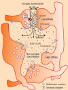
Non-synaptic receptors and transporters involved in brain functions and targets of drug treatment
- PMID: 20136842
- PMCID: PMC2935987
- DOI: 10.1111/j.1476-5381.2009.00624.x
Non-synaptic receptors and transporters involved in brain functions and targets of drug treatment
Abstract
Beyond direct synaptic communication, neurons are able to talk to each other without making synapses. They are able to send chemical messages by means of diffusion to target cells via the extracellular space, provided that the target neurons are equipped with high-affinity receptors. While synaptic transmission is responsible for the 'what' of brain function, the 'how' of brain function (mood, attention, level of arousal, general excitability, etc.) is mainly controlled non-synaptically using the extracellular space as communication channel. It is principally the 'how' that can be modulated by medicine. In this paper, we discuss different forms of non-synaptic transmission, localized spillover of synaptic transmitters, local presynaptic modulation and tonic influence of ambient transmitter levels on the activity of vast neuronal populations. We consider different aspects of non-synaptic transmission, such as synaptic-extrasynaptic receptor trafficking, neuron-glia communication and retrograde signalling. We review structural and functional aspects of non-synaptic transmission, including (i) anatomical arrangement of non-synaptic release sites, receptors and transporters, (ii) intravesicular, intra- and extracellular concentrations of neurotransmitters, as well as the spatiotemporal pattern of transmitter diffusion. We propose that an effective general strategy for efficient pharmacological intervention could include the identification of specific non-synaptic targets and the subsequent development of selective pharmacological tools to influence them.
Figures


Similar articles
-
Glia and volume transmission during physiological and pathological states.J Neural Transm (Vienna). 2005 Jan;112(1):137-47. doi: 10.1007/s00702-004-0120-4. Epub 2004 Mar 19. J Neural Transm (Vienna). 2005. PMID: 15599612 Review.
-
Non-synaptic interaction between neurons in the brain, an analog system: far from Cajal-Sherringtons's galaxy.Bull Mem Acad R Med Belg. 2003;158(10-12):373-9; discussion 379-80. Bull Mem Acad R Med Belg. 2003. PMID: 15244343
-
Characterization of a novel tonic gamma-aminobutyric acidA receptor-mediated inhibition in magnocellular neurosecretory neurons and its modulation by glia.Endocrinology. 2006 Aug;147(8):3746-60. doi: 10.1210/en.2006-0218. Epub 2006 May 4. Endocrinology. 2006. PMID: 16675519
-
Modulatory role of presynaptic nicotinic receptors in synaptic and non-synaptic chemical communication in the central nervous system.Brain Res Brain Res Rev. 1999 Nov;30(3):219-35. doi: 10.1016/s0165-0173(99)00016-8. Brain Res Brain Res Rev. 1999. PMID: 10567725 Review.
-
Glia-neuron intercommunications and synaptic plasticity.Prog Neurobiol. 1996 Jun;49(3):185-214. doi: 10.1016/s0301-0082(96)00012-3. Prog Neurobiol. 1996. PMID: 8878303 Review.
Cited by
-
Presynaptic Release-Regulating mGlu1 Receptors in Central Nervous System.Front Pharmacol. 2016 Aug 31;7:295. doi: 10.3389/fphar.2016.00295. eCollection 2016. Front Pharmacol. 2016. PMID: 27630571 Free PMC article. Review.
-
How cigarette smoking may increase the risk of anxiety symptoms and anxiety disorders: a critical review of biological pathways.Brain Behav. 2013 May;3(3):302-26. doi: 10.1002/brb3.137. Epub 2013 Mar 26. Brain Behav. 2013. PMID: 23785661 Free PMC article.
-
Intercellular signaling pathway among Endothelia, astrocytes and neurons in excitatory neuronal damage.Int J Mol Sci. 2013 Apr 16;14(4):8345-57. doi: 10.3390/ijms14048345. Int J Mol Sci. 2013. PMID: 23591846 Free PMC article. Review.
-
Intrinsic organization of the corpus callosum.Front Physiol. 2024 Jul 1;15:1393000. doi: 10.3389/fphys.2024.1393000. eCollection 2024. Front Physiol. 2024. PMID: 39035452 Free PMC article. Review.
-
Constitutive and Conditional Epitope Tagging of Endogenous G-Protein-Coupled Receptors in Drosophila.J Neurosci. 2024 Aug 14;44(33):e2377232024. doi: 10.1523/JNEUROSCI.2377-23.2024. J Neurosci. 2024. PMID: 38937100 Free PMC article.
References
-
- Abbracchio MP, Burnstock G, Verkhratsky A, Zimmermann H. Purinergic signalling in the nervous system: an overview. Trends Neurosci. 2009;32:19–29. - PubMed
-
- Abercrombie ED, Keller RW, Jr, Zigmond MJ. Characterization of hippocampal norepinephrine release as measured by microdialysis perfusion: pharmacological and behavioral studies. Neuroscience. 1988;27:897–904. - PubMed
-
- Agnati LF, Fuxe K, Zoli M, Ozini I, Toffano G, Ferraguti F. A correlation analysis of the regional distribution of central enkephalin and beta-endorphin immunoreactive terminals and of opiate receptors in adult and old male rats. Evidence for the existence of two main types of communication in the central nervous system: the volume transmission and the wiring transmission. Acta Physiol Scand. 1986;128:201–207. - PubMed
-
- Agnati LF, Zoli M, Stromberg I, Fuxe K. Intercellular communication in the brain: wiring versus volume transmission. Neuroscience. 1995;69:711–726. - PubMed
Publication types
MeSH terms
Substances
LinkOut - more resources
Full Text Sources
Medical

