. 2010 Mar;13(2):279-88.
doi: 10.1111/j.1467-7687.2009.00883.x.

Sensori-motor experience leads to changes in visual processing in the developing brain

Affiliations

Sensori-motor experience leads to changes in visual processing in the developing brain

Karin Harman James. Dev Sci. 2010 Mar.

Abstract

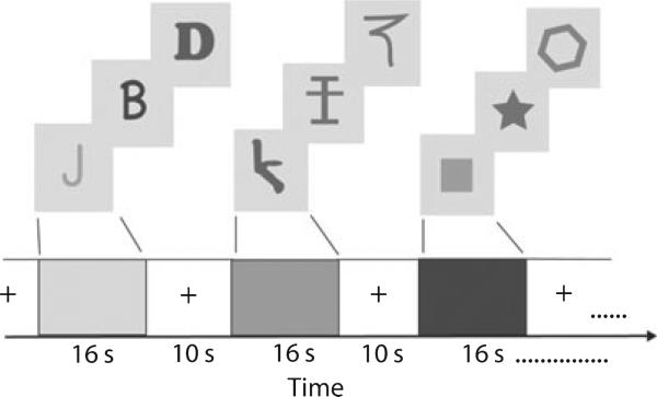
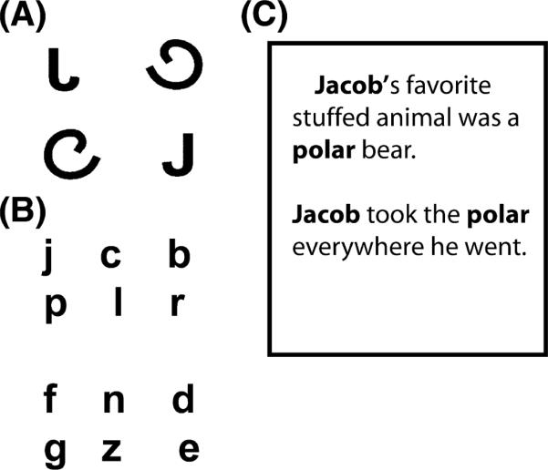
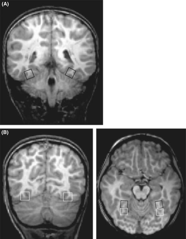
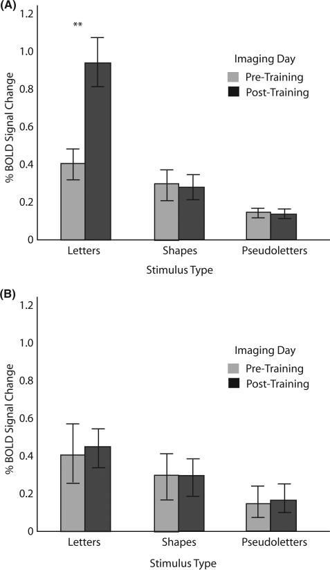
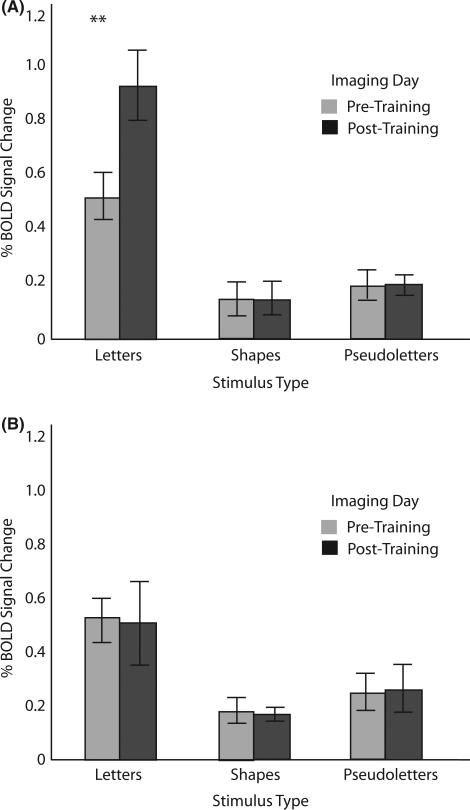
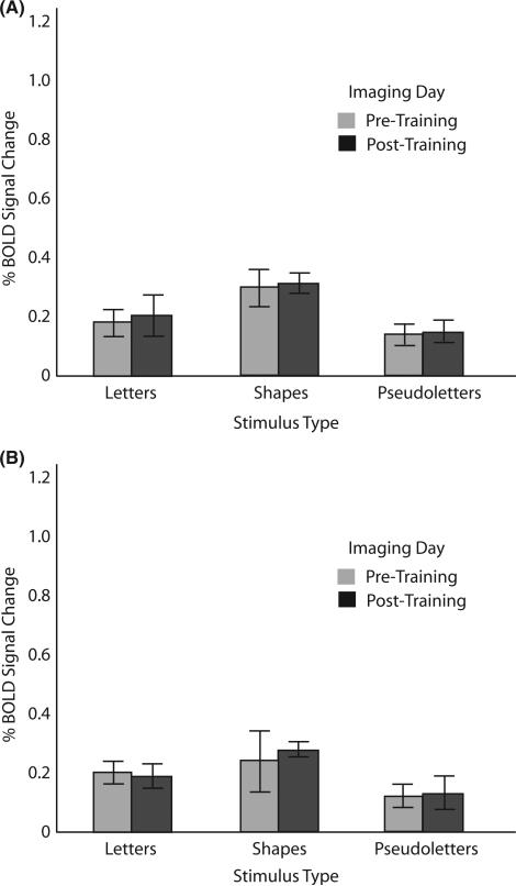
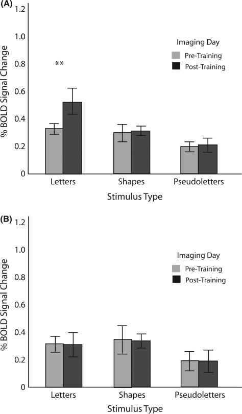
Since Broca's studies on language processing, cortical functional specialization has been considered to be integral to efficient neural processing. A fundamental question in cognitive neuroscience concerns the type of learning that is required for functional specialization to develop. To address this issue with respect to the development of neural specialization for letters, we used functional magnetic resonance imaging (fMRI) to compare brain activation patterns in pre-school children before and after different letter-learning conditions: a sensori-motor group practised printing letters during the learning phase, while the control group practised visual recognition. Results demonstrated an overall left-hemisphere bias for processing letters in these pre-literate participants, but, more interestingly, showed enhanced blood oxygen-level-dependent activation in the visual association cortex during letter perception only after sensori-motor (printing) learning. It is concluded that sensori-motor experience augments processing in the visual system of pre-school children. The change of activation in these neural circuits provides important evidence that 'learning-by-doing' can lay the foundation for, and potentially strengthen, the neural systems used for visual letter recognition.


Figures

Figure 1
Figure 1
Graphical depiction of fMRI design and stimuli used in the present study (see text for design details).
Figure 2
Figure 2
(A) An example of the four-alternative forced-choice task: participants were asked to ‘point to the “J”. (B) Words that contained the letters to be studied were highlighted in the story text. Participants were given examples in both upper and lower case. (C) The letters were presented in this format on a piece of paper: participants then either named or wrote the letters with feedback.
Figure 3
Figure 3
(A) Anatomical locale of the anterior fusiform gyrus presented on a coronal slice. (B) anatomical locale of the posterior fusiform presented on a coronal slice, left, and both the anterior and posterior fusiform presented on the transverse slice. Because these are not transformed into Talairach space, coordinates are not provided.
Figure 4
Figure 4
Left posterior fusiform region of interest. (A) Percentage blood oxygen-level-dependent (BOLD) signal change as a function of stimulus type during pre-training and post-training imaging sessions for the sensori-motor training group. (B) Percentage BOLD signal change as a function of stimulus type during pre-training and post-training imaging sessions for the visual-only training group. On this and all other graphs error bars represent standard error of the mean; ** depict significant differences at p < .01; and * depict differences at p < .05.
Figure 5
Figure 5
Left anterior fusiform gyrus. (A) Percentage blood oxygen-level-dependent (BOLD) signal change as a function of stimulus type during pre-training and post-training imaging sessions for the sensori-motor training group. (B) Percentage BOLD signal change as a function of stimulus type during pre-training and post-training imaging sessions for the visual-only training group.
Figure 6
Figure 6
Right posterior fusiform gyrus. (A) Percentage blood oxygen-level-dependent (BOLD) signal change as a function of stimulus type during pre-training and post-training imaging sessions for the sensori-motor training group. (B) Percentage BOLD signal change as a function of stimulus type during pre-training and post-training imaging sessions for the visual-only training group.
Figure 7
Figure 7
Right anterior fusiform gyrus. (A) Percentage blood oxygen-level-dependent (BOLD) signal change as a function of stimulus type during pre-training and post-training imaging sessions for the sensori-motor training group. (B) Percentage BOLD signal change as a function of stimulus type during pre-training and post-training imaging sessions for the visual-only training group.

References

    1. Bader LA. Bader Reading and Language inventory. Merrill Prentice Hall; New York: 2005.
    1. Brooks R. Intelligence without representation. Artificial Intelligence Journal. 1991a;47:139–160.
    1. Casey BJ, Tottenham N, Liston C, Durston S. Imaging the developing brain: what have we learned about cognitive development? Trends in Cognitive Science. 2005;9(3):104–110. - PubMed
    1. Chao LL, Martin A. Representation of man-made objects in the dorsal stream. Neuroimage. 2000;12:478–484. - PubMed
    1. Clark A. Being there: Putting brain body and world together again. MIT Press; Cambridge, MA: 1997.

Publication types

LinkOut - more resources