Review
. 2010 Feb;22(1):55-62.
doi: 10.1016/j.coi.2010.01.003.

Infection and apoptosis as a combined inflammatory trigger

Affiliations
Review

Infection and apoptosis as a combined inflammatory trigger

Miriam Beer Torchinsky et al. Curr Opin Immunol. 2010 Feb.

Abstract

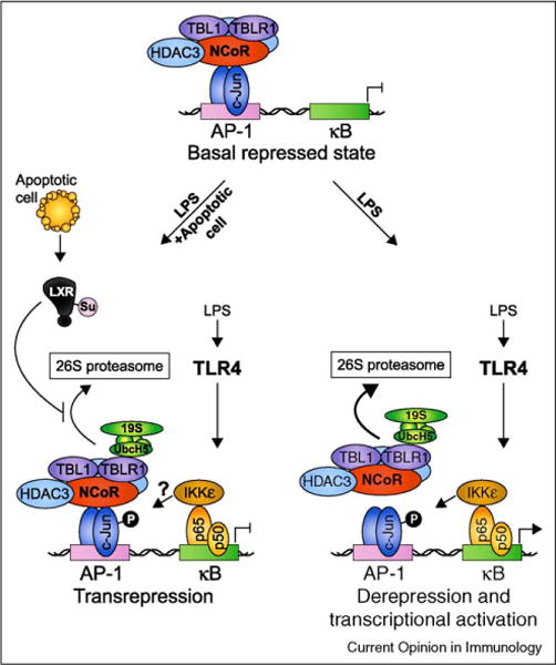
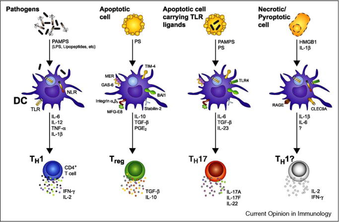
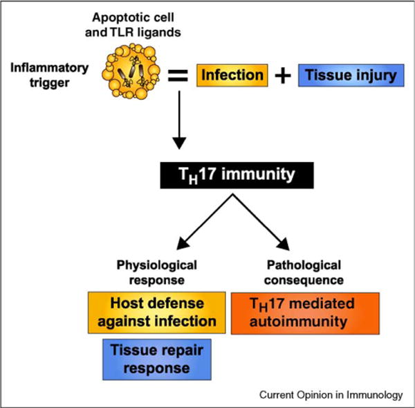
While inflammatory phagocytosis of microbial pathogens and non-inflammatory phagocytosis of apoptotic cells have each been studied extensively, the consequences of innate immune recognition of host cells undergoing apoptosis as a direct result of infection are unclear. In this situation, the innate immune system is confronted with mixed signals, those from apoptotic cells and those from the infecting pathogen. Nuclear receptor activation has been implicated downstream of apoptotic cell recognition while Toll-like receptors are the prototypical inflammatory receptors engaged during infection. When the two signals combine, a new set of events takes place beginning with transrepression of a subset of inflammatory-response genes and ending with the induction of a T helper-17 adaptive immune response. This response is best suited for clearing the infecting pathogen and repairing the damage that occurred to the host tissue during infection.


Figures

Figure 1
Figure 1
Ligand-dependent transrepression as a possible mechanism for apoptotic cell-mediated suppression of TLR-induced inflammatory gene expression. In the repressed ‘off’ state, LPS-induced genes such as the well characterized iNOS gene are actively suppressed by a multisubunit complex containing NCoR, HDAC3, TBL1, and TBLR1. When TLR4 signaling is initiated, IκB is degraded and NF-κB p50-p65 subunits enter the nucleus and bind to κB elements in the promoter. Phosphorylation of p65 on S536 creates a docking site for IκB Kinase ε (IKKε) allowing it to phosphorylate adjacent c-Jun/NCoR complexes and initiate corepressor clearance through TBLR1-mediated recruitment of UbcH5 and the 19S proteasome, ubiquitination, and subsequent degradation by the 26S proteasome. Binding of NF-κB allows recruitment of coactivators resulting in derepression and transcriptional activation. In this way, coupled AP-1/κB elements have been suggested to serve as ‘integrated circuits’ for switching promoters from a repressed to an activated state in response to an inflammatory trigger [28••]. When both TLR ligands and nuclear receptor ligands are present (depicted here as ‘LPS + apoptotic cell’ where the ligands for LXR are derived from apoptotic cells), ligand binding to LXR results in its SUMOylation by the E2 ligase Ubc9 and the E3 ligase HDAC4 and subsequent ability to interfere with NCoR corepressor clearance [27]. The result is ligand-dependent transrepression of inflammatory-response gene transcription.
Figure 2
Figure 2
While phagocytosis of TLR ligand-carrying apoptotic cells instructs TH17 differentiation, innate recognition of other forms of cell death may have different immune consequences. DC facing an infection sense a multitude of PAMPs expressed by microbes via a variety of PRRs such as TLR and NLRs. DC undergo a maturation program leading to the expression of T-cell co-stimulatory molecules and the secretion of inflammatory cytokines including IL-6, IL-12, IL-1β, and TNF-α. IL-12 production is the main driver of CD4+ T-cell differentiation into TH1 [6]. By contrast, when DC phagocytose an apoptotic cell, they induce a non-inflammatory response associated with the differentiation of Treg cells. Cells that succumb to apoptosis express ‘eat-me’ signals including exposure of PS at the outer leaflet of the plasma membrane. PS is subsequently recognized by DC through several receptors such as TIM-4, stabilin-2, BAI1, MER, and the αvβ3 integrin. While TIM-4, stabilin-2, and BAI1 recognize PS directly, MER or the αvβ3 integrin require interaction with bridging molecules such as GAS6 or MFG-E8, respectively, to recognize PS and initiate phagocytosis. DC can then prime CD4+ T cells to differentiate into regulatory T cells by secreting anti-inflammatory cytokines such as TGF-β and IL-10. In the particular case of an apoptotic cell carrying TLR ligands, for example a neutrophil undergoing apoptosis following phagocytosis of bacteria, the combination of pro-inflammatory signals from activated TLRs with signals driven by the apoptotic cell cargo induces a unique cocktail of cytokines including IL-6, TGF-β, and IL-23, and triggers TH17 differentiation. Other types of cell death such as necrosis and pyroptosis also exist and these are thought to be immunogenic because of the release of cellular contents and inflammatory cytokines such as IL-1β that can activate DC [49]. While HMGB1 released by necrotic cells is known to result in DC maturation when recognized by RAGE, the ligand for CLEC9A, a C-type lectin receptor implicated in recognition of necrotic cells, remains to be discovered [49]. The consequence of phagocytosis of necrotic or pyroptotic cells on CD4+ T-cell differentiation is not known. HMGB1, high-mobility group box 1; RAGE, receptor for advanced glycation end-products; CLEC9A, C-type lectin domain family 9, member A.
Figure 3
Figure 3
Proposed physiological and pathological outcomes of innate recognition of apoptotic cells dying from infection. When cells undergo apoptosis as a result of infection, the inflammatory trigger presented to the innate immune system is that of both infection and tissue injury. While PAMPs trigger inflammatory PRRs such as TLRs, apoptotic ‘eat-me’ signals trigger anti-inflammatory signaling pathways. Given that apoptosis frequently occurs during infection, the ability of the immune system to mount an effective response against pathogens despite the presence of immunosuppressive dying cells has been a long-standing paradox. A TH17 adaptive immune response reconciles the tolerance induced by apoptotic cell clearance with the necessarily inflammatory nature of infections. Through the release of inflammatory and reparative cytokines, TH17 cells can initiate not only defense against the pathogen, but also repair of injured tissues. Because one of the signals that trigger TH17 immunity consists of dying host cells, the pathological consequences of innate recognition of apoptotic cells during infection could be the development of autoimmunity.

References

    1. Vaux DL, Korsmeyer SJ. Cell death in development. Cell. 1999;96:245–254. - PubMed
    1. Gaipl US, Munoz LE, Grossmayer G, Lauber K, Franz S, Sarter K, Voll RE, Winkler T, Kuhn A, Kalden J, et al. Clearance deficiency and systemic lupus erythematosus (SLE) J Autoimmun. 2007;28:114–121. This paper showed that patients with SLE had apoptotic cell material in dendritic cells located in lymph node germinal centers, and autoantibodies that had gained autoreactivity in germinal center reactions. This supported the hypothesis that impaired clearance of apoptotic cells in patients with SLE could lead to secondary necrosis and buildup of self-antigens in tissues, contributing to autoimmunity in this disease. - PubMed
    1. Janko C, Schorn C, Grossmayer GE, Frey B, Herrmann M, Gaipl US, Munoz LE. Inflammatory clearance of apoptotic remnants in systemic lupus erythematosus (SLE) Autoimmun Rev. 2008;8:9–12. - PubMed
    1. Blander JM, Medzhitov R. On regulation of phagosome maturation and antigen presentation. Nat Immunol. 2006;7:1029–1035. - PubMed
    1. Kinchen JM, Ravichandran KS. Phagosome maturation: going through the acid test. Nat Rev Mol Cell Biol. 2008;9:781–795. - PMC - PubMed

Publication types