. 2010 Feb 23;20(4):339-45.
doi: 10.1016/j.cub.2009.12.035. Epub 2010 Feb 4.

The actin-bundling protein fascin stabilizes actin in invadopodia and potentiates protrusive invasion

Affiliations

The actin-bundling protein fascin stabilizes actin in invadopodia and potentiates protrusive invasion

Ang Li et al. Curr Biol. .

Abstract

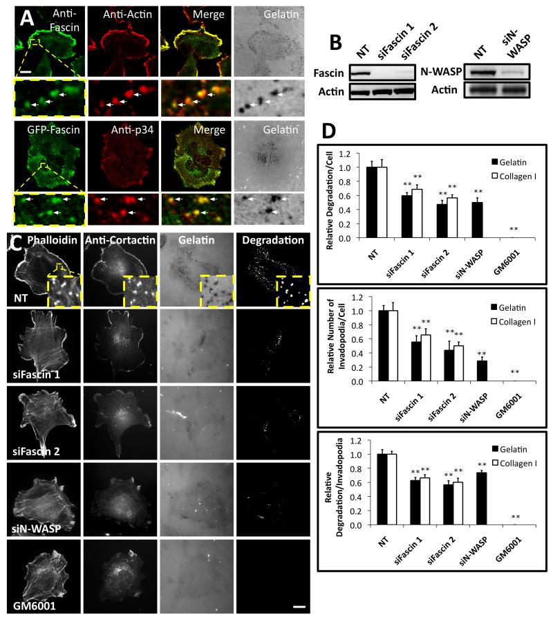
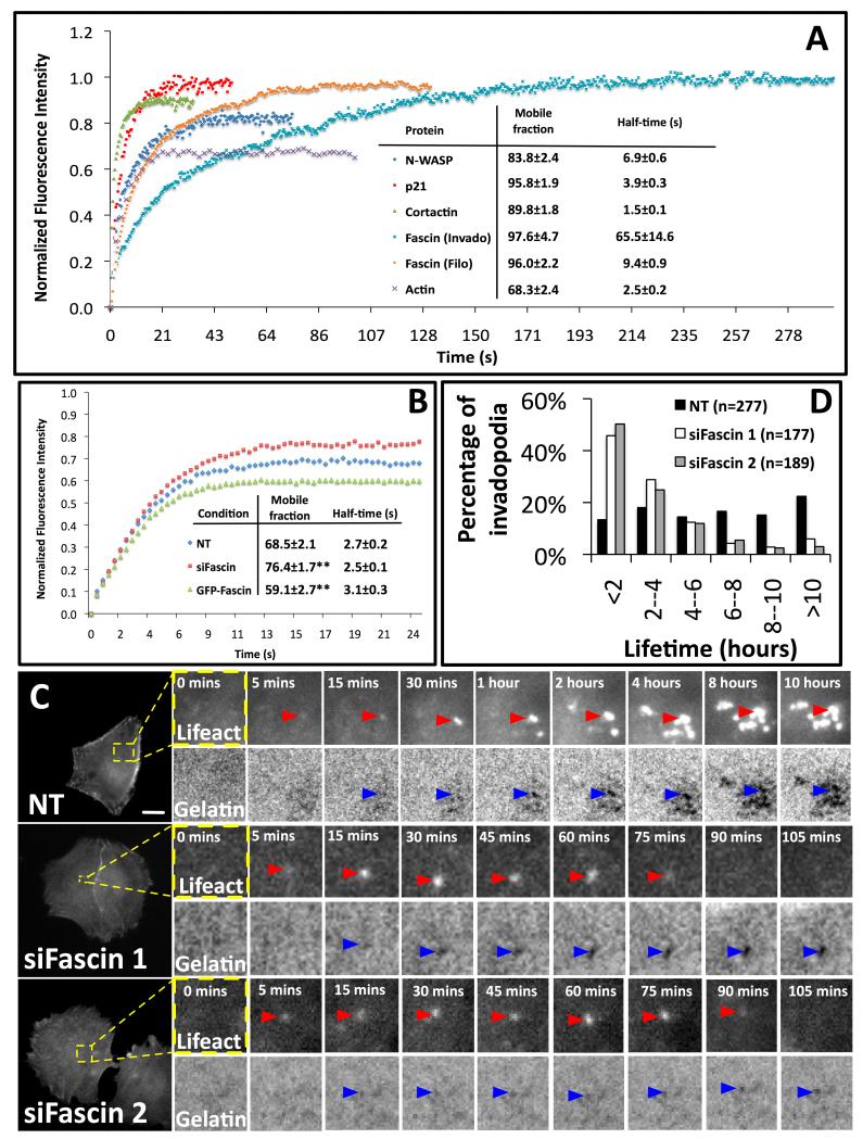
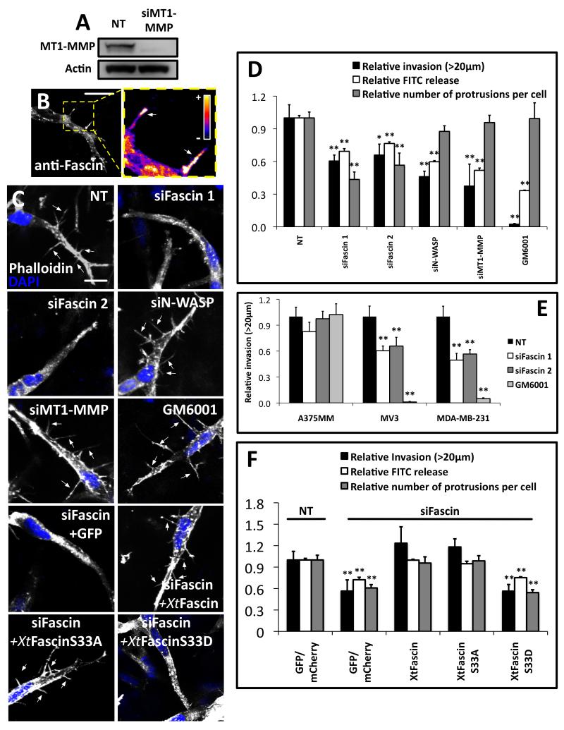
Fascin is an actin-bundling protein involved in filopodia assembly and cancer invasion and metastasis of multiple epithelial cancer types. Fascin forms stable actin bundles with slow dissociation kinetics in vitro and is regulated by phosphorylation of serine 39 by protein kinase C (PKC). Cancer cells use invasive finger-like protrusions termed invadopodia to invade into and degrade extracellular matrix. Invadopodia have highly dynamic actin that is assembled by both Arp2/3 complex and formins; they also contain components of membrane trafficking machinery such as dynamin and cortactin and have been compared with focal adhesions and podosomes. We show that fascin is an integral component of invadopodia and that it is important for the stability of actin in invadopodia. The phosphorylation state of fascin at S39, a PKC site, contributes to its regulation at invadopodia. We further implicate fascin in invasive migration into collagen I-Matrigel gels and particularly in cell types that use an elongated mesenchymal type of motility in 3D. We provide a potential molecular mechanism for how fascin increases the invasiveness of cancer cells, and we compare invadopodia with invasive filopod-like structures in 3D.


Figures

Figure 1
Figure 1. Fascin is important for invadopodia assembly and matrix degradation
(A) Top, CHL-1 human melanoma cells on Oregon Green® 488 gelatin matrix (grey) with anti-fascin (primary 55K2) and anti-actin. Bottom, cells expressing GFP-fascin on Alexa594 gelatin matrix (grey) with anti-p34-Arc (ARPC2). Arrows- invadopodia. Bar, 10μm. (B) Western blots of lysates from CHL-1 cells with anti-fascin (Left), anti-N-WASP (Right) or anti-actin (Loading control, left and right) (C) CHL-1 cells expressing siRNAs as indicated or treated with 5 μM GM6001 on Oregon Green® 488 gelatin matrix with rhodamine phalloidin (F-actin) and anti-cortactin. Bars, 10μm. (D) Top: Relative area of degradation per cell on gelatin (Black) and Collagen I (White). Middle: number of invadopodia per cell on gelatin (Black) and Collagen I (White). Bottom: area of degradation per invadopodium of the ten biggest degradations per cell on gelatin (Black) and Collagen I (White). All error bars indicate (Mean ± SEM). **, P< 0.01 by t-test. Invadopodia were defined as puncta enriched for actin, cortactin and gelatin degradation. Similar experiments done with A375MM or MDA-MB231 cells are shown in Figure S1. Detailed analysis of fascin localization at invadopodia is shown in Figure S2. Movie 1 shows dynamics of actin comets at invadopodia and 3D reconstruction of fascin localization in comets.
Figure 2
Figure 2. Fascin regulation at serine 39 is important for invaopodia formation
(A) CHL-1 cells stably expressing GFP, GFP-X.tropicalis Fascin, GFP-X. tropicalis Fascin S33A and GFP-X. tropicalis Fascin S33D were transfected with NT control siRNA or Fascin siRNA (siFascin 1), cultured on Alexa594 gelatin matrix and labeled with anti-cortactin. Arrows- invadopodia. Bars, 10μm (B) Western blot showing stable expression of GFP-X.tropicalis Fascin or mutants following fascin knockdown in CHL-1 cells (C) Top: Relative area of degradation on gelatin per cell (as in Figure 1). Middle: area of degradation per invadopod (as in Figure 1). Bottom: number of invadopodia per cell (as in Figure 1). Invadopodia were defined as cortactin puncta colocalizing with area of matrix degradation and error bars show (Mean ± SEM). **, P< 0.01 by t-test. See Figure S3 for related data with A375MM cells.
Figure 3
Figure 3. Fascin is stably associated with invadopodia and promotes long lifetime
(A) The recovery kinetics of GFP-cortactin (green triangles) (n=28), GFP-actin (purple crosses) (n=31), GFP-p21-Arc (red squares) (n=30), GFP-N-WASP (blue diamond) (n=30) at invadopodia and GFP-fascin at invadopodia (light blue x) (n=22) and filopodia (orange dots) (n=25) after photobleaching. Mobile fraction and half time of recovery are indicated. (Mean ± SEM). (B) Recovery kinetics of mRFP-actin at invadopodia in cells expressing NT control siRNA (blue diamond) (n=50), GFP-fascin and control siRNA (EGFP-fascin) (green triangles) (n=47) or fascin siRNA (siFascin) (red squares) (n=45). Percentage mobile fraction and half time of recovery for each condition are indicated. (Mean ± SEM) **, P< 0.01 by t-test. (C) Timelapse image sequence of A375MM cells stably expressing GFP-Lifeact expressing control or fascin siRNA on gelatin matrix. Bars, 10μm. Red arrowheads - invadopodia and blue arrowheads - gelatin degradation. D, Time-lapse lifetime of invadopodia was calculated with data from 30 cells from at least three independent experiments. (n=the total number of invadopodia analyzed). See Figure S4 and Movies 2 and 3 for photobleaching and timelapse of invadopodia lifetime.
Figure 4
Figure 4. Fascin is required for efficient mesenchymal type invasion in 3D
(A) Western blot of CHL-1 cells expressing NT control siRNA, MT1-MMP siRNA probed with anti-MT1-MMP and anti-actin loading control. (B) Left, CHL-1 in collagen I-Matrigel matrix fixed and stained with anti-fascin (55K2). Serial z-stack images (0.5 μm) were combined and shown here. Bars, 10μm. Right, densitometric analysis of fascin signal with arrows at peak fascin intensities (C) CHL-1 cells in 3D collagen I-Matrigel matrix treated with siRNA, inhibitor or rescued with Xtfascin constructs (as indicated) were fixed and stained with rhodamine phalloidin and DAPI. Serial z-stack images (0.5 μm interval) were combined. Bars, 10μm. Filopod-like protrusions were arrowed. (D) Relative invasion >20μM into collagen I-Matrigel (Black); relative release of soluble FITC from collagenolysis caused by CHL-1 cells within 3D FITC–collagen lattices (White) and relative number of filopod-like protrusions per cell (Grey). (E) Summaries of invasion assays of A375MM, MV3 and MDA-MB-231 cells treated with NT, Fascin siRNA or 5μM GM6001 using collagen I-Matrigel matrix. (F) Relative invasion into 3D collagen I-Matrigel, relative release of soluble FITC from collagenolysis caused by cells within 3D FITC–collagen lattices (White) and relative number of filopod-like protrusions per cell (Grey) in cells expressing GFP and NT siRNA, GFP and siFascin, or XtFascin and mutants as indicated on the figure. All error bars show (Mean ± SEM). **, P< 0.01; *, P<0.05 by t-test. Relative invasion and filopod-like protrusions were quantified as described in Supplementary Experimental Procedures. Figure S4C shows the localization of GFP-MT1-MMP to filopod-like protrusions in 3D collagen I-Matrigel matrix. Movie S4 shows the dynamics of filopod-like protrusions in 3D and a comparison of NT and fascin knockdown cells.

Similar articles

Cited by

References

    1. Tseng Y, Fedorov E, McCaffery JM, Almo SC, Wirtz D. Micromechanics and ultrastructure of actin filament networks crosslinked by human fascin: a comparison with alpha-actinin. J Mol Biol. 2001;310:351–366. - PubMed
    1. Adams JC. Roles of fascin in cell adhesion and motility. Curr Opin Cell Biol. 2004;16:590–596. - PubMed
    1. Baldassarre M, Ayala I, Beznoussenko G, Giacchetti G, Machesky LM, Luini A, Buccione R. Actin dynamics at sites of extracellular matrix degradation. Eur J Cell Biol. 2006;85:1217–1231. - PubMed
    1. Lizarraga F, Poincloux R, Romao M, Montagnac G, Le Dez G, Bonne I, Rigaill G, Raposo G, Chavrier P. Diaphanous-related formins are required for invadopodia formation and invasion of breast tumor cells. Cancer Res. 2009;69:2792–2800. - PubMed
    1. Buccione R, Caldieri G, Ayala I. Invadopodia: specialized tumor cell structures for the focal degradation of the extracellular matrix. Cancer Metastasis Rev. 2009;28:137–149. - PubMed

Publication types