Review
. 2010 Apr;20(4):214-22.
doi: 10.1016/j.tcb.2010.01.003.

NuMA after 30 years: the matrix revisited

Affiliations
Review

NuMA after 30 years: the matrix revisited

Andreea E Radulescu et al. Trends Cell Biol. 2010 Apr.

Abstract

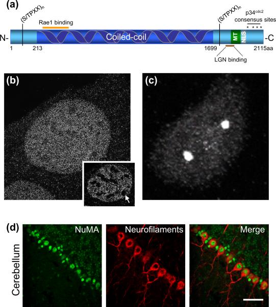
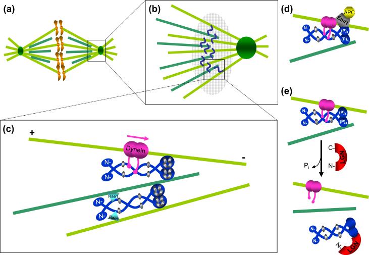
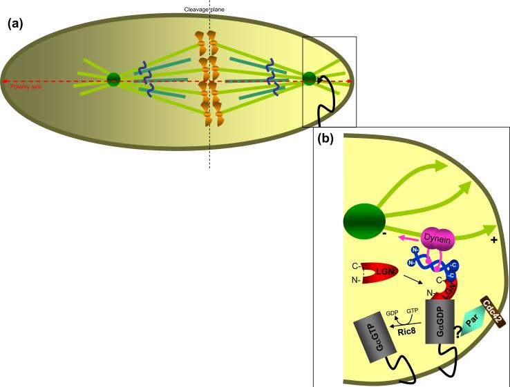
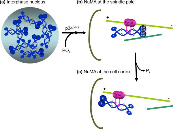
The large nuclear mitotic apparatus (NuMA) protein is an abundant component of interphase nuclei and an essential player in mitotic spindle assembly and maintenance. With its partner, cytoplasmic dynein, NuMA uses its cross-linking properties to tether microtubules to spindle poles. NuMA and its invertebrate homologs play a similar tethering role at the cell cortex, thereby mediating essential asymmetric divisions during development. Despite its maintenance as a nuclear component for decades after the final mitosis of many cell types (including neurons), an interphase role for NuMA remains to be established, although its structural properties implicate it as a component of a nuclear scaffold, perhaps as a central constituent of the proposed nuclear matrix.


Figures

Figure 1
Figure 1
NuMA is a component of the nucleus and the spindle poles. (a) Schematic representation of human NuMA protein domains. Dark blue, coiled coil domain; light blue, N- and C- globular termini; gray, nuclear localization signal (NLS), residues 1971–1991 [7]; green (MT), microtubule binding domain, residues 1900–1971 [7]; brown line, LGN binding sequence (1878–1910 [7]); orange line, Rae1 binding domain, residues 325–829 [38]. S/TPXX, a DNA-binding motif found in gene regulatory proteins, occurs six times in the N- and seven times in the C- termini, respectively, of NuMA [8]. Asterisks, four p34cdc2 phosphorylation sites (threonine, 2000, 2040, 2091, serine 2072) [23]. (b) NuMA appears to fill up interphase nuclei of immortalized mouse embryonic fibroblasts. Single sections through the nucleus (inset) reveal exclusion domains (arrow). Images were acquired using structured illumination microscopy, on an OMX microscope (UC, Davis). Unpublished data. (c) NuMA is enriched at the spindle poles of mitotic HeLa cells (image acquired using an Olympus FV1000 spectral deconvolution confocal microscope). Unpublished data. (d) NuMA is expressed in nuclei of post-mitotic neurons. Immunofluorescence staining of frozen cerebellar sections from an adult C57/B6 mouse, with antibodies against NuMA (green) and unphosphorylated neurofilaments (red). Images in (d), courtesy of Dr. Alain Silk, unpublished data. Scale bar, 50μm.
Figure 2
Figure 2
NuMA functions at spindle poles to bundle and tether microtubules. (a) Schematic of the mitotic spindle, representing kinetochore (light green) and non-kinetochore (dark green) microtubules. The spindle pole (square box) is enlarged in (b). NuMA (blue squiggle) cross-links kinetochore and non-kinetochore microtubules at the spindle pole. NuMA containing complexes involved in microtubule linkage at the spindle poles form a structure equivalent to a “spindle pole matrix” (large hatched oval). (c) Proteins interacting with NuMA at spindle poles (enlarged square box from (b)). During nuclear breakdown, NuMA is phosphorylated (yellow) at four putative p34cdc2 sites (threonine 2000, 2040, 2091, serine 2072) [23, 25]. Mitotically phosphorylated NuMA associates with dynein (pink), which carries it in a minus-end directed fashion (pink arrow) and deposits it at spindle poles where it functions in spindle maintenance via microtubule tethering [20]. A NuMA dimer (dark blue, top) cross-links microtubules through interactions mediated by its C-terminal microtubule binding domain and the associated dynein (pink) [6, 7]. Rae1, a messenger RNA transport protein (blue triangle), may also mediate binding of the NuMA N-termini to microtubules [38], thus transiently generating a higher level of parallel microtubule bundling (bottom microtubules). (d) Emi1 (gray), an inhibitor of Cdc20 dependent activation of the anaphase promoting complex (APC), has been proposed to exist in a complex with dynein and NuMA prior to anaphase [28], as a means of sequestering APCCdc20 (yellow) away from the general cytoplasm. (e) A possible mechanism for NuMA dissociation from microtubules. LGN (red), by competing with microtubules for NuMA binding, acts as a negative regulator of NuMA bundling [7] and may be responsible for the relocalization of a NuMA subset from the spindle pole to the cell cortex.
Figure 3
Figure 3
Spindle positioning and asymmetric cell division. (a) During asymmetric cell division the mitotic spindle is positioned along the polarity axis (red dashed horizontal line) and tethered to one pole of the cell (square box), thus establishing an off center cleavage plane (dashed vertical line). (b) Protein complexes involved in spindle orientation (enlarged square box from (a)). External cues, an adherens junctions/ Cdc42 complex (brown) and Par (light blue) determine mother cell polarity [86, 87]. The Par complex recruits the GDP-bound, myristoylated Gα subunit of the trimeric G protein, Gi, and this interaction is mediated by a poorly described protein complex [44]. Normally, LGN exists in the cytoplasm in a closed conformation (red, unbound). Dual binding of NuMA and Gα at its C- and N-termini, respectively, switches LGN to an open conformation (red, bound). These interactions ensure recruitment of NuMA to specific sites at the cell cortex [42]. Ric8 (Resistance to inhibitors of cholinesterase 8) is a guanine nucleotide exchange factor (GEF) for Gαi-GDP [88, 89]. The Ric8-mediated release of Gαi-GTP results in dissociation of the LGN/ NuMA/ Gα complex.
Figure 4
Figure 4

Similar articles

Cited by

  • Force transduction by cadherin adhesions in morphogenesis.
    Pannekoek WJ, de Rooij J, Gloerich M. Pannekoek WJ, et al. F1000Res. 2019 Jul 10;8:F1000 Faculty Rev-1044. doi: 10.12688/f1000research.18779.1. eCollection 2019. F1000Res. 2019. PMID: 31327995 Free PMC article. Review.
  • Poly(ADP-ribose) polymerase 3 (PARP3), a newcomer in cellular response to DNA damage and mitotic progression.
    Boehler C, Gauthier LR, Mortusewicz O, Biard DS, Saliou JM, Bresson A, Sanglier-Cianferani S, Smith S, Schreiber V, Boussin F, Dantzer F. Boehler C, et al. Proc Natl Acad Sci U S A. 2011 Feb 15;108(7):2783-8. doi: 10.1073/pnas.1016574108. Epub 2011 Jan 26. Proc Natl Acad Sci U S A. 2011. PMID: 21270334 Free PMC article.
  • Differential proteomic analysis of mammalian tissues using SILAM.
    McClatchy DB, Liao L, Park SK, Xu T, Lu B, Yates Iii JR. McClatchy DB, et al. PLoS One. 2011 Jan 20;6(1):e16039. doi: 10.1371/journal.pone.0016039. PLoS One. 2011. PMID: 21283754 Free PMC article.
  • The Global Phosphorylation Landscape of SARS-CoV-2 Infection.
    Bouhaddou M, Memon D, Meyer B, White KM, Rezelj VV, Correa Marrero M, Polacco BJ, Melnyk JE, Ulferts S, Kaake RM, Batra J, Richards AL, Stevenson E, Gordon DE, Rojc A, Obernier K, Fabius JM, Soucheray M, Miorin L, Moreno E, Koh C, Tran QD, Hardy A, Robinot R, Vallet T, Nilsson-Payant BE, Hernandez-Armenta C, Dunham A, Weigang S, Knerr J, Modak M, Quintero D, Zhou Y, Dugourd A, Valdeolivas A, Patil T, Li Q, Hüttenhain R, Cakir M, Muralidharan M, Kim M, Jang G, Tutuncuoglu B, Hiatt J, Guo JZ, Xu J, Bouhaddou S, Mathy CJP, Gaulton A, Manners EJ, Félix E, Shi Y, Goff M, Lim JK, McBride T, O'Neal MC, Cai Y, Chang JCJ, Broadhurst DJ, Klippsten S, De Wit E, Leach AR, Kortemme T, Shoichet B, Ott M, Saez-Rodriguez J, tenOever BR, Mullins RD, Fischer ER, Kochs G, Grosse R, García-Sastre A, Vignuzzi M, Johnson JR, Shokat KM, Swaney DL, Beltrao P, Krogan NJ. Bouhaddou M, et al. Cell. 2020 Aug 6;182(3):685-712.e19. doi: 10.1016/j.cell.2020.06.034. Epub 2020 Jun 28. Cell. 2020. PMID: 32645325 Free PMC article.
  • Polar relaxation by dynein-mediated removal of cortical myosin II.
    Chapa-Y-Lazo B, Hamanaka M, Wray A, Balasubramanian MK, Mishima M. Chapa-Y-Lazo B, et al. J Cell Biol. 2020 Aug 3;219(8):e201903080. doi: 10.1083/jcb.201903080. J Cell Biol. 2020. PMID: 32497213 Free PMC article.

References

    1. Lydersen BK, Pettijohn DE. Human-specific nuclear protein that associates with the polar region of the mitotic apparatus: distribution in a human/hamster hybrid cell. Cell. 1980;22:489–499. - PubMed
    1. Compton DA, et al. Primary structure of NuMA, an intranuclear protein that defines a novel pathway for segregation of proteins at mitosis. J Cell Biol. 1992;116:1395–1408. - PMC - PubMed
    1. Kallajoki M, et al. Microinjection of a monoclonal antibody against SPN antigen, now identified by peptide sequences as the NuMA protein, induces micronuclei in PtK2 cells. J Cell Sci. 1993;104(Pt 1):139–150. - PubMed
    1. Yang CH, et al. NuMA: an unusually long coiled-coil related protein in the mammalian nucleus. J Cell Biol. 1992;116:1303–1317. - PMC - PubMed
    1. Gueth-Hallonet C, et al. NuMA: a bipartite nuclear location signal and other functional properties of the tail domain. Exp Cell Res. 1996;225:207–218. - PubMed