. 2010 Apr 4;9(4):403-13.
doi: 10.1016/j.dnarep.2009.12.019. Epub 2010 Feb 4.

RecQ-dependent death-by-recombination in cells lacking RecG and UvrD

Affiliations

RecQ-dependent death-by-recombination in cells lacking RecG and UvrD

Natalie C Fonville et al. DNA Repair (Amst). .

Abstract

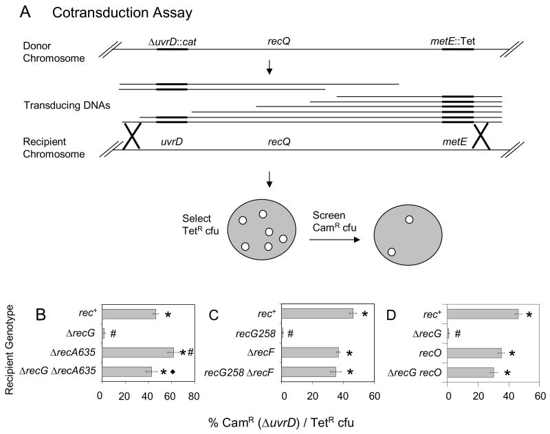
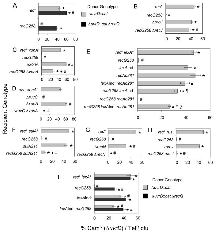
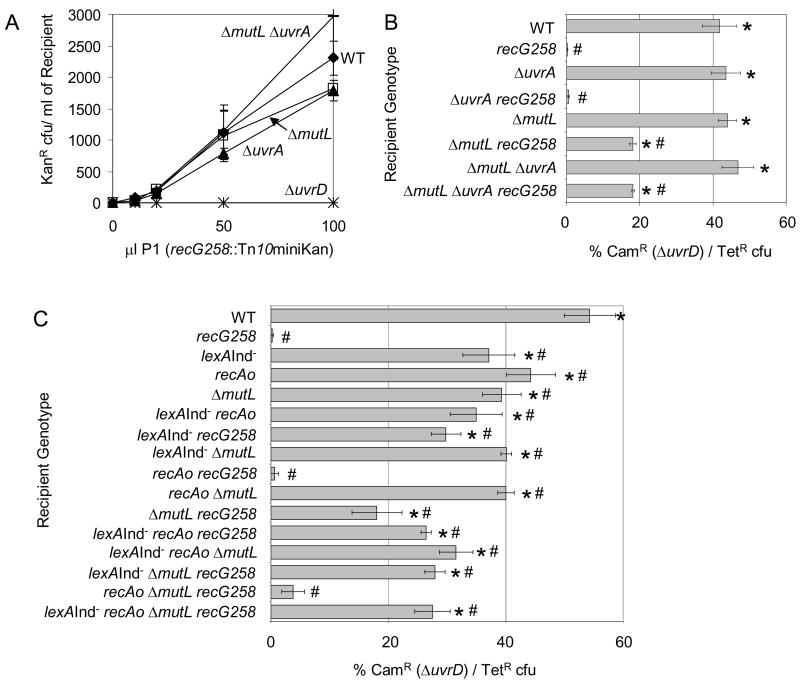
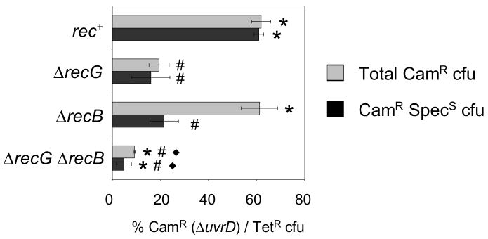
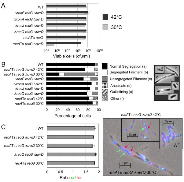
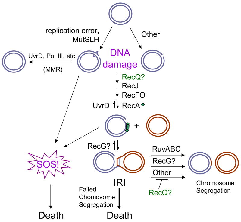
Maintenance of genomic stability is critical for all cells. Homologous recombination (HR) pathways promote genome stability using evolutionarily conserved proteins such as RecA, SSB, and RecQ, the Escherichia coli homologue of five human proteins at least three of which suppress genome instability and cancer. A previous report indicated that RecQ promotes the net accumulation in cells of intermolecular HR intermediates (IRIs), a net effect opposite that of the yeast and two human RecQ homologues. Here we extend those conclusions. We demonstrate that cells that lack both UvrD, an inhibitor of RecA-mediated strand exchange, and RecG, a DNA helicase implicated in IRI resolution, are inviable. We show that the uvrD recG cells die a "death-by-recombination" in which IRIs accumulate blocking chromosome segregation. First, their death requires RecA HR protein. Second, the death is accompanied by cytogenetically visible failure to segregate chromosomes. Third, FISH analyses show that the unsegregated chromosomes have completed replication, supporting the hypothesis that unresolved IRIs prevented the segregation. Fourth, we show that RecQ and induction of the SOS response are required for the accumulation of replicated, unsegregated chromosomes and death, as are RecF, RecO, and RecJ. ExoI exonuclease and MutL mismatch-repair protein are partially required. This set of genes is similar but not identical to those that promote death-by-recombination of DeltauvrD Deltaruv cells. The data support models in which RecQ promotes the net accumulation in cells of IRIs and RecG promotes resolution of IRIs that form via pathways not wholly identical to those that produce the IRIs resolved by RuvABC. This implies that RecG resolves intermediates other than or in addition to standard Holliday junctions resolved by RuvABC. The role of RecQ in net accumulation of IRIs may be shared by one or more of its human homologues.


Figures

Fig. 1
Fig. 1
Demonstration of inviability of recG ΔuvrD double mutants by cotransduction and requirements for RecA, RecF and RecO for the inviability. (A) Cotransduction assay. Phage P1 grown on a donor strain with two linked antibiotic resistances (SMR9811) recombine into a recipient strain. Transductants are selected for the non-lethal marker (TetR) and screened for those that also contain the potentially lethal marker (ΔuvrD::cat). recQ is located between uvrD and the linked Tet marker, so for examining a requirement for RecQ (Fig. 2) P1 grown on SMR9812, a strain that is ΔrecQ, is used. (B) ΔrecG ΔuvrD cells are inviable and RecA is required for their inviability. ΔuvrD cannot be cotransduced into a ΔrecG recipient (SMR10698), but can into rec+ (SMR10424), ΔrecA (SMR10427) and ΔrecA ΔrecG (SMR10428) recipients carrying pML104-3. (C) RecF is required for recG ΔuvrD inviability. ΔuvrD cannot be cotransduced into recG258 (SMR10419), but can into rec+ (SMR6319), ΔrecF (SMR10423) and ΔrecF recG258 (SMR10437) recipients. (D) RecO is required for ΔrecG ΔuvrD inviability. ΔuvrD cannot be cotransduced into ΔrecG (SMR11133), but can into recO (SMR11134) and recO ΔrecG (SMR11135) recipients. Mean ± SEM of 3 experiments for B, C and D. * indicates a significant difference from recG (B–D). # indicates a significant difference from rec+ (B–D); ◆ indicates a significant difference from the ΔrecA single mutant (B, p = 0.02) of the double mutant indicated.
Fig. 2
Fig. 2
Roles of recombination proteins and the SOS response in the death of recG uvrD cells. Data are results of co-transduction experiments as described in Fig. 1. (A) RecQ is required for recG ΔuvrD inviability. ΔuvrD and ΔrecQ alleles can be cotransduced together into recG258 recipient cells (SMR10419, donor strain SMR9812), but the ΔuvrD mutation alone (donor SMR9811) cannot. rec+ positive-control recipient, SMR6319. (B) RecJ is required for recG ΔuvrD inviability. ΔuvrD can be cotransduced into both ΔrecJ (SMR10434) and ΔrecJ recG258 (SMR10439) recipients. (C) ExoI is partially required for recG ΔuvrD inviability. ΔuvrD can be cotransduced into a ΔxonA recipient (SMR10395) efficiently, and a ΔxonA recG258 recipient (SMR10438) with intermediate efficiency. (D) ExoI is not required for inviability of ΔruvC ΔuvrD cells. ΔuvrD cannot be cotransduced into a ΔxonA ΔruvC recipient (SMR10417). ΔxonA positive control, SMR10395; ruvC negative control, SMR10408. (E) The SOS response, SOS-induced levels of RecA, and induction of another SOS gene(s) promote recG ΔuvrD inviability. Efficient cotransduction of ΔuvrD into “SOS-off” lexA(Ind) (SMR9801), recAo281 (SMR9809) cells which produce SOS-induced levels of RecA, lexA(Ind) recAo281 (SMR9805), and lexA(Ind) recG258 (SMR10440) recipients, indicates that shutting off the SOS response via the lexA(Ind) mutation partially restores viability to recG ΔuvrD cultures. Cotransduction efficiency is partially reduced from the level in lexA(Ind) recG258 in a lexA(Ind) recAo281 recG258 recipient (SMR10441), but is not abolished as in recAo281 recG258 (SMR10442) and recG258 (SMR10419). Therefore, SOS-induced levels of RecA account for some but not all of the contribution of the SOS response to recG ΔuvrD inviability. (F) SulA contributes little to recG ΔuvrD inviability. ΔuvrD is cotransduced poorly into a sulA211 recG258 recipient (SMR10443). sulA211 positive control, SMR9837. (G) RecN is not required for recG ΔuvrD inviability. ΔuvrD cannot be cotransduced into a ΔrecN recG258 recipient (SMR10734). ΔrecN positive control, SMR10730. (H) Activation of expression of Rus endonuclease by the rus-1 mutation does not restore viability to recG ΔuvrD cells. ΔuvrD cannot be cotransduced into recG258 rus-1 recipients (SMR10746). rec+ rus+ control, SMR10743; rus-1 positive control, SMR10744; recG258 negative control, SMR10745. (I) RecQ promotes recG ΔuvrD inviability wholly or partly independently of induction of the SOS response. ΔuvrD and ΔrecQ alleles confer greater viability when cotransduced together into recG258 lexA(Ind) recipient cells (SMR10440, donor strain SMR9812) than the ΔuvrD alone (donor SMR9811), implying that ΔrecQ promotes recG ΔuvrD inviability wholly or partly independently of the LexA/SOS response. Mean ± SEM of 3 experiments (A–I). * indicates a significant difference from recG; # indicates a significant difference from rec+ (A–I). ◆ indicates a significant difference of each double mutant tested from the following isogenic single mutant: ΔxonA (C, D), sulA (F), recN (G), and rus-1 (H). In (A) recG cotransduced with ΔuvrD ΔrecQ is significantly different from rec+ cotransduced with same. In (E), the § indicated strain is significantly different from all others in the panal while the ¶ indicated strain is significantly different from the lexA(Ind) single mutant among mutants carrying the lexA(Ind) allele. In (I) lexA(Ind) recG cotransduced with ΔuvrD ΔrecQ is significantly different from the same strain cotransduced with ΔuvrD alone. It is also significantly different from recG cotransduced with the ΔrecQ ΔuvrD donor.
Fig. 3
Fig. 3
Incomplete mismatch repair and not nucleotide excision repair contributes to death of recG ΔuvrD cells. (A) Simple loss of either MMR or NER does not cause inviability of recG cells, in that quantitative transduction of recG258 (SMR9932 donor) into strains mutant for proteins involved in NER (ΔuvrA, SMR8977), MMR (ΔmutL, SMR8982) or both (ΔuvrA ΔmutL, SMR8986) is not impaired relative to MMR- and NER-proficient cells (SMR6319). Therefore, blocking formation of the MMR and NER intermediates created by MutL and UvrA does not create an inviability with recG. ΔuvrD negative control, SMR8976. (B) Although simple loss of either MMR or NER does not cause inviability of recG cells, loss of the MutL step in MMR relieves some of the inviability of recG ΔuvrD cultures. This implies that MMR intermediates initiated by MutL can be lethal to recG cells if UvrD is not present to complete repair and remove those intermediates. ΔuvrD can be cotransduced with intermediate efficiency into a ΔmutL recG258 recipient (SMR11116) indicating a partial requirement for MutL, but not UvrA, in the death of recG ΔuvrD cultures. recG258 SMR10419; ΔuvrA recG258 SMR11115; MMR- and NER-proficient cells SMR6319 (“WT”); ΔuvrA SMR8977; ΔmutL SMR8982; ΔuvrA ΔmutL SMR8986; and ΔmutL ΔuvrA recG258 SMR11117 recipient cells. (C) The combination of incomplete MMR and SOS-induction does not account for all of the recG ΔuvrD inviability. When combined, the partial requirement for MutL and the SOS response does not restore more viability to recG ΔuvrD cells than lexA(Ind) or ΔmutL alone, or in the presence of SOS-induced levels of RecA. MMR- and SOS-proficient cells SMR6319; recG258 SMR10419; lexA(Ind) SMR9801; recAo281 SMR9809; ΔmutL SMR11323; lexA(Ind) recAo281 SMR9805; lexA(Ind) recG258 SMR10440; lexA(Ind) ΔmutL SMR11318; recAo281 recG258 SMR10442; recAo281 ΔmutL SMR11320; ΔmutL recG258 SMR11116; lexA(Ind) recAo281 recG258 SMR10441; lexA(Ind) recAo281 ΔmutL SMR11319; lexA(Ind) ΔmutL recG258 SMR11325; recAo281 ΔmutL recG258 SMR11329; lexA(Ind) recAo281 ΔmutL recG258 SMR11327. Mean ± SEM of 3 experiments (A–C). * indicates a significant difference from recG; # indicates a significant difference from WT (B, C). In (B) the triple ΔmutL ΔuvrA recG258 mutant is significantly different from all of the constituent single and double mutants except for ΔmutL recG258 (p= 0.99). In (C) the quadruple mutant lexA(Ind) recAo281 ΔmutL recG258 is significantly different from recAo281; lexA(Ind) ΔmutL; recAo281 recG258; recAo281 ΔmutL (p= 0.01); ΔmutL (p= 0.02), and recAo281 ΔmutL recG258. For WT compared to recAo, p= 0.02.
Fig. 4
Fig. 4
RecB is not required for death of ΔrecG ΔuvrD cells. ΔuvrD cannot be cotransduced into ΔrecG (SMR11188) or ΔrecB ΔrecG (SMR11190), but can into rec+ (SMR10424) and ΔrecB (SMR11189). All strains carry pML104-3 therefore all TetR cfu were also tested for SpecR to assay the loss of pML104-3 at the restrictive temperature, 37°C. Note that the somewhat higher cotransduction into recG cells seen here is an apparent effect of the altered temperature regiment with higher temperature allowing greater viability (data not shown). Mean ± SEM of 3 experiments. * indicates a significant difference from recG, # indicates a significant difference from WT, and ◆ indicates a significant difference from ΔrecB. For ΔrecG compared to ΔrecG ΔrecB p ≤ 0.05.
Fig. 5
Fig. 5
Chromosome-segregation defects but completed replication during synchronous death of recG ΔuvrD cells. (A) Upon shift of recA(Ts) recG ΔuvrD cells (SMR10740) switched to the permissive temperature (30°C, RecA+ phenotype), from the restrictive temperature (42°C, RecA phenotype), cells die synchronously and show (B) chromosome-segregation defects as indicated by an increased percentage of filamented cells with unsegregated nucleoids. The chromosome-segregation defect is also rescued by the additional mutation of ΔrecF, ΔxonA, ΔrecJ, or ΔrecQ (Strains SMR10735, SMR10736, SMR10737 and SMR10738 respectively). Control recA(Ts) recG cells (SMR10739) do not show chromosome-segregation defects at the permissive temperature. Mean of 3 experiments. SEM is < 5% of cells/category for all genotypes and temps. (C) Completion of chromosome replication in dying recA(Ts) recG ΔuvrD cells shown by fluorescence in situ hybridization (FISH) to ori (green foci) and terminus-proximal (red foci) chromosomal sequences. Left, the ratios of ori:ter foci in cells are unchanged during synchronous death of recA(Ts) recG ΔuvrD (SMR10740) cells at permissive or restrictive temperatures from those of the non-dying wild-type (rec+, SMR6319) control. The ratio of ori:ter is not different from that of rec+ or recA(Ts) recG (SMR10739) cells, or of ΔrecQ recG ΔuvrD (SMR10738) cells. Right, representative example of FISH data. Images are overlays of phase contrast (blue), red (ter hybridization) and green (ori hybridization) exposures. Red arrows, examples of red ter foci; green arrows, examples of green ori foci. Mean ± SEM of 3 experiments for B and C; ≥ 500 cells scored/genotype/temperature/experiment.
Fig. 6
Fig. 6
Models for death of ΔrecG ΔuvrD cells. DNA damage arises from replication errors (mismatches) which are recognized and processed by MutSLH or from other sources. In the presence of UvrD, MMR can be completed to give intact dsDNA. In the absence of UvrD, the damaged DNA is processed by homologous recombination proteins. RecQ and RecJ may function together at this stage to create ssDNA gaps which are substrates for RecF, O and RecA, which can promote both the induction of SOS and the initiation of recombination (formation of IRIs). IRIs are then resolved in multiple pathways, primarily through the action of RuvABC, but also via the action of RecG. Both the persistence of unresolved IRIs and the SOS response can lead to loss of viability (death.) RecG might branch migrate D-loops or IRIs to contribute to their elimination. RecQ might either promote the formation of ssDNA required for IRI formation or inhibit an alternative resolution pathway to Ruv and RecG. Blue and Red circles represent dsDNA.

Similar articles

Cited by

References

    1. Bohr VA. Rising from the RecQ-age: the role of human RecQ helicases in genome maintenance. Trends Biochem Sci. 2008;33:609–620. - PMC - PubMed
    1. Hu Y, Raynard S, Sehorn MG, Lu X, Bussen W, Zheng L, Stark JM, Barnes EL, Chi P, Janscak P, Jasin M, Vogel H, Sung P, Luo G. RECQL5/Recql5 helicase regulates homologous recombination and suppresses tumor formation via disruption of Rad51 presynaptic filaments. Genes Dev. 2007;21:3073–3084. - PMC - PubMed
    1. Sharma S, Brosh RM., Jr Unique and important consequences of RECQ1 deficiency in mammalian cells. Cell Cycle. 2008;7:989–1000. - PMC - PubMed
    1. Friedberg E, Walker G, Siede W, Wood R, Schultz R, Ellenberger T. DNA Repair and Mutagenesis. 2. ASM Press; Washington, D.C: 2005.
    1. Mimitou EP, Symington LS. Nucleases and helicases take center stage in homologous recombination. Trends Biochem Sci. 2009;34:264–272. - PubMed

Publication types

MeSH terms

LinkOut - more resources