

Self-complementary AAV5 vector facilitates quicker transgene expression in photoreceptor and retinal pigment epithelial cells of normal mouse
- PMID: 20138034
- PMCID: PMC2854182
- DOI: 10.1016/j.exer.2010.01.011
Self-complementary AAV5 vector facilitates quicker transgene expression in photoreceptor and retinal pigment epithelial cells of normal mouse
Abstract
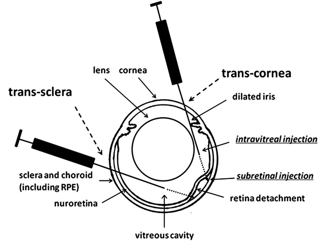
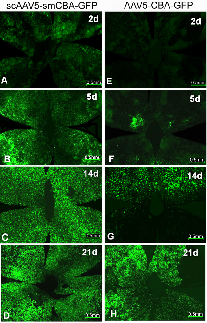
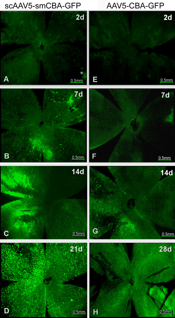
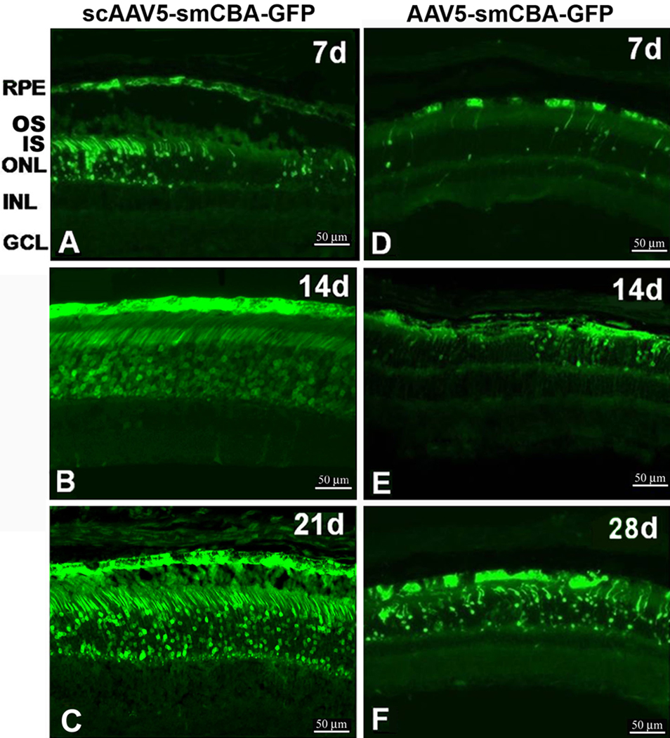
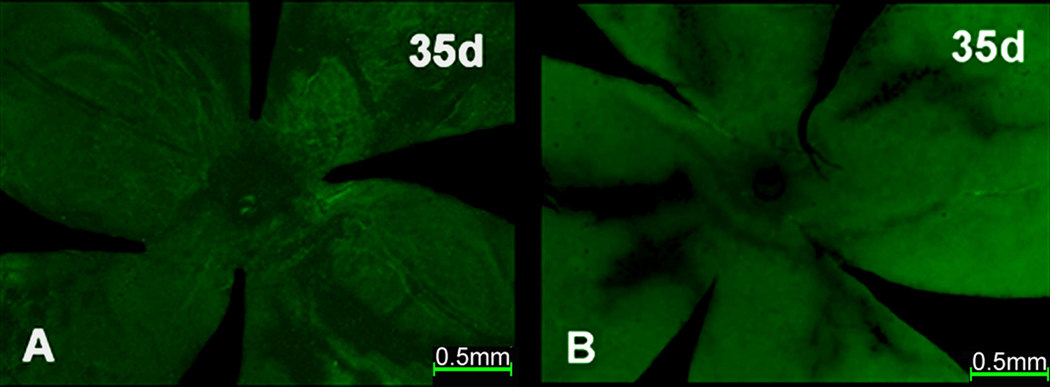
To clarify whether transduction efficiency and cell type specificity of self-complementary (sc) AAV5 vectors are similar to those of standard, single-stranded AAV5 vectors in normal retina, one micro liter of scAAV5-smCBA-GFP vector (1 x 10(12) genome-containing particles/ml) and AAV5-smCBA-GFP vector (1 x 10(12) genome-containing particles/ml) were subretinally or intravitreally (in both cases through the cornea) injected into the right and left eyes of adult C57BL/6J mice, respectively. On post-injection day (PID) 1, 2, 5, 7, 10, 14, 21, 28 and 35, eyes were enucleated; retinal pigment epithelium (RPE) wholemounts, neuroretinal wholemounts and eyecup sections were prepared to evaluate green fluorescent protein (GFP) expression by fluorescent microscopy. GFP expression following trans-cornea subretinal injection of scAAV5-smCBA-GFP vector was first detected in RPE wholemounts around PID 1 and in neuroretinal wholemounts between PID 2 and 5; GFP expression peaked and stabilized between PID 10-14 in RPE wholemounts and between P14 and P21 in neuroretinal wholemounts with strong, homogeneous green fluorescence covering the entire wholemounts. The frozen sections supported the following findings from the wholemounts: GFP expression appeared first in RPE around PID 1-2 and soon spread to photoreceptors (PR) cells; by PID 7, moderate GFP expression was found mainly in PR and RPE layers; between PID 14 and 21, strong and homogenous GFP expression was observed in RPE and PR cells. GFP expression following subretinal injection of AAV5-smCBA-GFP was first detected in RPE wholemounts around PID 5-7 and in neuroretinal wholemounts around PID 7-10; ssAAV5-mediated GFP expression peaked at PID 21 in RPE wholemounts and around PID 28 in neuroretinal wholemounts; sections from AAV5 treated eyes also supported findings obtained from wholemounts: GFP expression was first detected in RPE and then spread to the PR cells. Peak GFP expression in RPE mediated by scAAV5 was similar to that mediated by AAV5. However, peak GFP expression mediated by scAAV5 in PR cells was stronger than that mediated by AAV5. No GFP fluorescence was detected in any retinal cells (RPE wholemounts, neuroretinal wholemounts and retinal sections) after trans-cornea intravitreal delivery of either scAAV5-GFP or AAV5-GFP. Neither scAAV5 nor AAV5 can transduce retinal cells following trans-cornea intravitreal injection. The scAAV5 vector used in this study directs an earlier onset of transgene expression than the matched AAV5 vector, and has stronger transgene expression in PR cells following subretinal injection. Our data confirm the previous reports that scAAV vectors have an earlier onset than the standard, single strand AAV vectors (Natkunarajah et al., 2008; Yokoi et al., 2007). scAAV5 vectors may be more useful than standard, single-stranded AAV vector when addressing certain RPE and/or PR cell-related models of retinal dystrophy, particularly for mouse models of human retinitis pigmentosa that require rapid and robust transgene expression to prevent early degeneration in PR cells.
(c) 2010 Elsevier Ltd. All rights reserved.
Figures
Similar articles
-
Gene therapy following subretinal AAV5 vector delivery is not affected by a previous intravitreal AAV5 vector administration in the partner eye.Mol Vis. 2009;15:267-75. Epub 2009 Feb 6. Mol Vis. 2009. PMID: 19190735 Free PMC article.
-
Efficiency of lentiviral transduction during development in normal and rd mice.Mol Vis. 2006 Jul 11;12:756-67. Mol Vis. 2006. PMID: 16862069
-
Ocular gene transfer with self-complementary AAV vectors.Invest Ophthalmol Vis Sci. 2007 Jul;48(7):3324-8. doi: 10.1167/iovs.06-1306. Invest Ophthalmol Vis Sci. 2007. PMID: 17591905
-
Quantifying transduction efficiencies of unmodified and tyrosine capsid mutant AAV vectors in vitro using two ocular cell lines.Mol Vis. 2011 Apr 29;17:1090-102. Mol Vis. 2011. PMID: 21552473 Free PMC article.
-
Human blue-opsin promoter preferentially targets reporter gene expression to rat s-cone photoreceptors.Invest Ophthalmol Vis Sci. 2006 Aug;47(8):3505-13. doi: 10.1167/iovs.05-1670. Invest Ophthalmol Vis Sci. 2006. PMID: 16877422
Cited by
-
Preclinical dose response study shows NR2E3 can attenuate retinal degeneration in the retinitis pigmentosa mouse model RhoP23H+/.Gene Ther. 2024 May;31(5-6):255-262. doi: 10.1038/s41434-024-00440-6. Epub 2024 Jan 26. Gene Ther. 2024. PMID: 38273095 Free PMC article.
-
The Degeneration and Apoptosis Patterns of Cone Photoreceptors in rd11 Mice.J Ophthalmol. 2017;2017:9721362. doi: 10.1155/2017/9721362. Epub 2017 Jan 12. J Ophthalmol. 2017. PMID: 28168050 Free PMC article.
-
Novel insights into gene therapy in the cornea.Exp Eye Res. 2021 Jan;202:108361. doi: 10.1016/j.exer.2020.108361. Epub 2020 Nov 16. Exp Eye Res. 2021. PMID: 33212142 Free PMC article. Review.
-
Gene therapy using self-complementary Y733F capsid mutant AAV2/8 restores vision in a model of early onset Leber congenital amaurosis.Hum Mol Genet. 2011 Dec 1;20(23):4569-81. doi: 10.1093/hmg/ddr391. Epub 2011 Aug 31. Hum Mol Genet. 2011. PMID: 21880665 Free PMC article.
-
Heparan Sulfate Binding Promotes Accumulation of Intravitreally Delivered Adeno-associated Viral Vectors at the Retina for Enhanced Transduction but Weakly Influences Tropism.J Virol. 2016 Oct 14;90(21):9878-9888. doi: 10.1128/JVI.01568-16. Print 2016 Nov 1. J Virol. 2016. PMID: 27558418 Free PMC article.
References
-
- Auricchio A, Kobinger G, Anand V, Hildinger M, O'Connor E, Maguire AM, Wilson JM, Bennett J. Exchange of surface proteins impacts on viral vector cellular specificity and transduction characteristics: the retina as a model. Hum. Mol. Genet. 2001;10:3075–3081. - PubMed
-
- Bainbridge JW, Mistry A, Schlichtenbrede FC, Smith A, Broderick C, DeAlwis M, Georgiadis A, Taylor PM, Squires M, Sethi C, Charteris D, Thrasher AJ, Sargan D, Ali RR. Stable rAAV-mediated transduction of rod and cone photoreceptors in the canine retina. Gene Ther. 2003;10:1336–1344. - PubMed
-
- Bainbridge JW, Tan MH, Ali RR. Gene therapy progress and prospects: the eye. Gene Ther. 2006;13(16):1191–1197. - PubMed
-
- Bainbridge JW, Smith AJ, Barker SS, Robbie S, Henderson R, Balaggan K, Viswanathan A, Holder GE, Stockman A, Petersen-Jones S, Bhattacharya SS, Thrasher AJ, Fitzke FW, Carter BJ, Rubin GS, Moore AT, Ali RR. Effect of gene therapy on visual function in Leber's congenital amaurosis. N. Engl. J. Med. 2008;358:2231–2239. - PubMed
Publication types
MeSH terms
Substances
Grants and funding
LinkOut - more resources
Full Text Sources
Other Literature Sources
Research Materials
