. 2010 Apr 2;397(3):740-51.
doi: 10.1016/j.jmb.2010.01.065. Epub 2010 Feb 4.

Two intramolecular isopeptide bonds are identified in the crystal structure of the Streptococcus gordonii SspB C-terminal domain

Affiliations

Two intramolecular isopeptide bonds are identified in the crystal structure of the Streptococcus gordonii SspB C-terminal domain

Nina Forsgren et al. J Mol Biol. .

Abstract

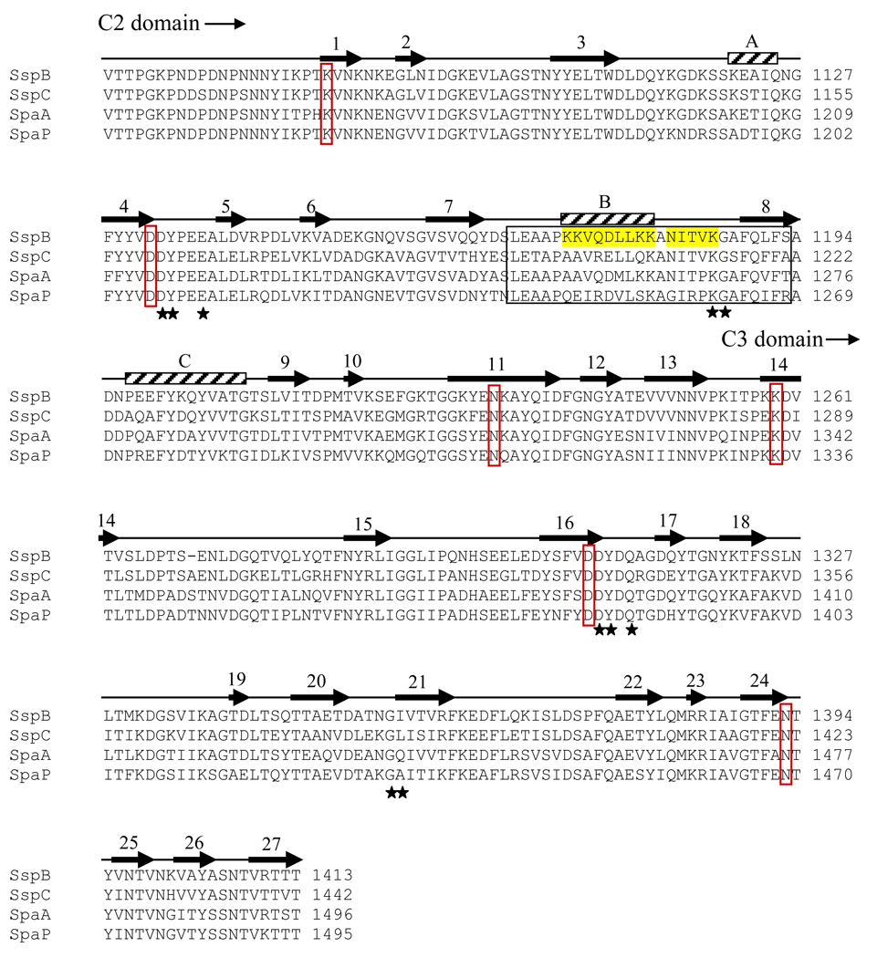
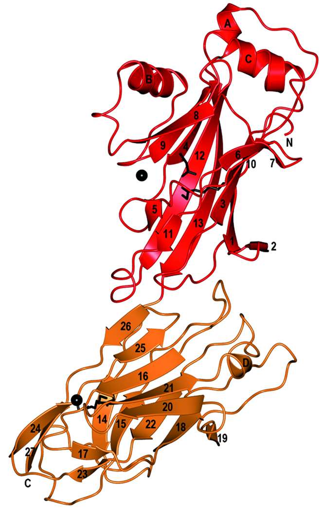
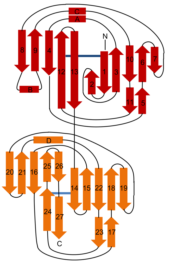
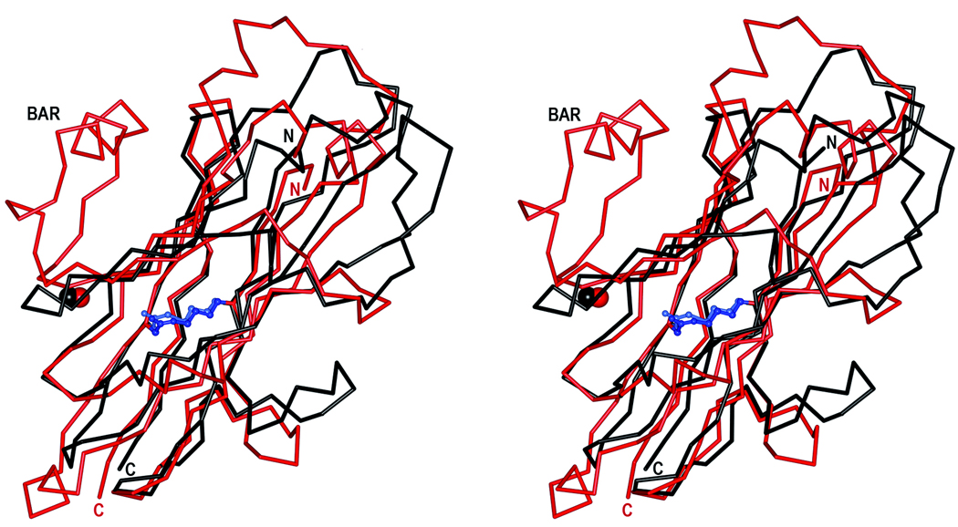
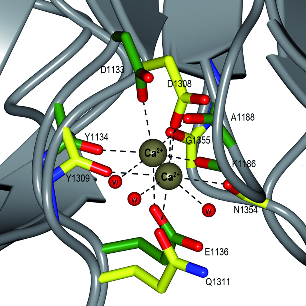
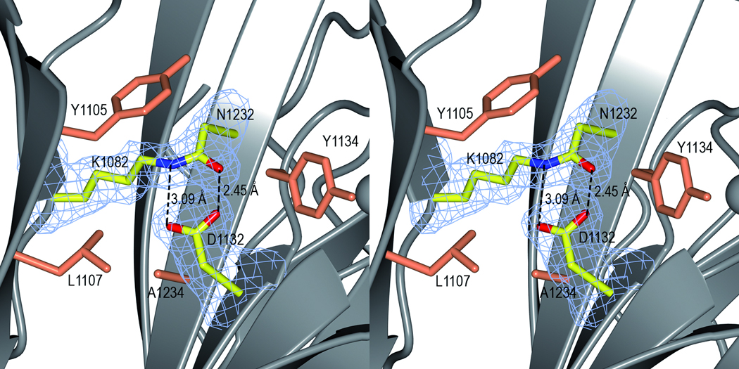
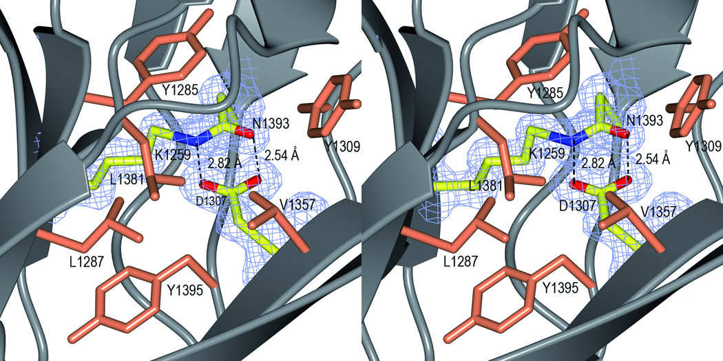
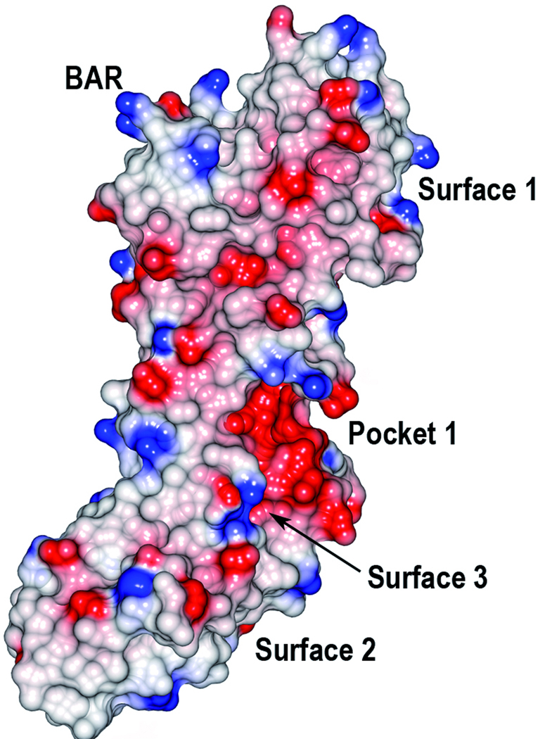
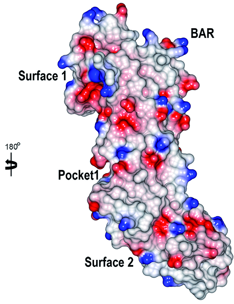
Streptococcus gordonii is a primary colonizer and is involved in the formation of dental plaque. This bacterium expresses several surface proteins. One of them is the adhesin SspB, which is a member of the Antigen I/II family of proteins. SspB is a large multi-domain protein that has interactions with surface molecules on other bacteria and on host cells, and is thus a key factor in the formation of biofilms. Here, we report the crystal structure of a truncated form of the SspB C-terminal domain, solved by single-wavelength anomalous dispersion to 1.5 A resolution. The structure represents the first of a C-terminal domain from a streptococcal Antigen I/II protein and is comprised of two structurally related beta-sandwich domains, C2 and C3, both with a Ca(2+) bound in equivalent positions. In each of the domains, a covalent isopeptide bond is observed between a lysine and an asparagine, a feature that is believed to be a common stabilization mechanism in Gram-positive surface proteins. S. gordonii biofilms contain attachment sites for the periodontal pathogen Porphyromonas gingivalis and the SspB C-terminal domain has been shown to have one such recognition motif, the SspB adherence region. The motif protrudes from the protein, and serves as a handle for attachment. The structure suggests several additional putative binding surfaces, and other binding clefts may be created when the full-length protein is folded.


Figures

Figure 1
Figure 1
Sequence alignment and overall structure of SspB-C1061–1413. a) Multiple sequence alignment of SspB-C1061–1413 and related AgI/II C-terminal domains. Sequences from the following streptococcal strains were compared; S. gordonii M5 (SspB), S. sanguinis SK36 (SspC), S. sobrinus MUCOB 263 (SpaA) and S. mutans UA159 (SpaP). Secondary structure elements of S.gordonii SspB-C1061–1413 are indicated above the sequence. The metal coordinating residues are marked with filled stars and residues involved in the isopeptide bonds are indicated by red boxes. The BAR region is boxed and the KKVQDLLKK and NITVK motifs are highlighted in yellow. b) Ribbon representation of SspB-C1061–1413. The C2 domain (residues1061-1253) is depicted in red and the C3 domain (residues 1254–1413) in orange. Ca2+ ions are depicted as black spheres. The isopeptide bonds between Lys 1082 and Asn1232 and between Lys1259 and Asn1393 are represented as stick models in black. c) Topology diagram of SspB-C1061–1413. β-strands are represented as arrows, helices as rectangles and loops as lines. Colouring as in (b).
Figure 1
Figure 1
Sequence alignment and overall structure of SspB-C1061–1413. a) Multiple sequence alignment of SspB-C1061–1413 and related AgI/II C-terminal domains. Sequences from the following streptococcal strains were compared; S. gordonii M5 (SspB), S. sanguinis SK36 (SspC), S. sobrinus MUCOB 263 (SpaA) and S. mutans UA159 (SpaP). Secondary structure elements of S.gordonii SspB-C1061–1413 are indicated above the sequence. The metal coordinating residues are marked with filled stars and residues involved in the isopeptide bonds are indicated by red boxes. The BAR region is boxed and the KKVQDLLKK and NITVK motifs are highlighted in yellow. b) Ribbon representation of SspB-C1061–1413. The C2 domain (residues1061-1253) is depicted in red and the C3 domain (residues 1254–1413) in orange. Ca2+ ions are depicted as black spheres. The isopeptide bonds between Lys 1082 and Asn1232 and between Lys1259 and Asn1393 are represented as stick models in black. c) Topology diagram of SspB-C1061–1413. β-strands are represented as arrows, helices as rectangles and loops as lines. Colouring as in (b).
Figure 2
Figure 2
Domain structure and stabilization. a) Superposition of the C2 and C3 domains. The C2 domain is coloured in red and the C3 domain in black. The isopeptide bonds are represented as ball-and-stick models in blue and light blue. The N-, and C-termini and the BAR motif are labelled. The picture is shown in stereo. b) Superposition of the metal binding sites in the C2 and C3 domains. The C2 residues are coloured in green and the C3 residues in yellow. The Ca2+ ions are depicted as brown spheres. The bonds involved the metal coordination are represented by dashed lines. c) The isopeptide bond in the C2 domain. The Lys-Asn isopeptide bond is represented as a stick model in a simulated annealing omit Fo-Fc map, contoured at 4σ. Hydrogen bonds to the aspartate residue are shown as dashed lines. Hydrophobic residues surrounding the isopeptide are shown in coral. The picture is shown in stereo. d) The isopeptide bond in the C3 domain. The figure is prepared as in c).
Figure 2
Figure 2
Domain structure and stabilization. a) Superposition of the C2 and C3 domains. The C2 domain is coloured in red and the C3 domain in black. The isopeptide bonds are represented as ball-and-stick models in blue and light blue. The N-, and C-termini and the BAR motif are labelled. The picture is shown in stereo. b) Superposition of the metal binding sites in the C2 and C3 domains. The C2 residues are coloured in green and the C3 residues in yellow. The Ca2+ ions are depicted as brown spheres. The bonds involved the metal coordination are represented by dashed lines. c) The isopeptide bond in the C2 domain. The Lys-Asn isopeptide bond is represented as a stick model in a simulated annealing omit Fo-Fc map, contoured at 4σ. Hydrogen bonds to the aspartate residue are shown as dashed lines. Hydrophobic residues surrounding the isopeptide are shown in coral. The picture is shown in stereo. d) The isopeptide bond in the C3 domain. The figure is prepared as in c).
Figure 3
Figure 3
Electrostatic surface of SspB-C1061–1413. The molecular surfaces are colored in red and blue according to positive and negative electrostatic potential, respectively. The two views are rotated 180° with respect to each other. The BAR motif, the putative binding pocket and surfaces are indicated.
Figure 3
Figure 3
Electrostatic surface of SspB-C1061–1413. The molecular surfaces are colored in red and blue according to positive and negative electrostatic potential, respectively. The two views are rotated 180° with respect to each other. The BAR motif, the putative binding pocket and surfaces are indicated.
Figure 4
Figure 4
Stereoview of the BAR handle. The BAR handle is important for recognition by the Porphyromonas gingivalis fimbriae Mfa1. The handle is constituted by two motifs; a helix (KKVQDLLKK, in yellow) followed by an extended region (NITVK, in orange). The side chains are represented as stick objects. A hydrogen bond between K1180 and N1126 in the β-sheet (grey) is marked with a dashed line. The position of the BAR handle is stabilized by a Ca2+ ion (black), coordinated by three main chain and two side chain oxygen atoms and a water molecule.

Similar articles

Cited by

References

    1. Jenkinson HF, Lamont RJ. Oral microbial communities in sickness and in health. Trends Microbiol. 2005;13:589–595. - PubMed
    1. Nyvad B, Kilian M. Comparison of the initial streptococcal microflora on dental enamel in caries-active and in caries-inactive individuals. Caries Res. 1990;24:267–272. - PubMed
    1. Jenkinson HF, Lamont RJ. Streptococcal adhesion and colonization. Crit. Rev. Oral Biol. Med. 1997;8:175–200. - PubMed
    1. Rosan B, Lamont RJ. Dental plaque formation. Microbes Infect. 2000;2:1599–1607. - PubMed
    1. Douglas CW, Heath J, Hampton KK, Preston FE. Identity of viridans streptococci isolated from cases of infective endocarditis. J. Med. Microbiol. 1993;39:179–182. - PubMed

Publication types