. 2010 May 1;48(9):1188-96.
doi: 10.1016/j.freeradbiomed.2010.01.039. Epub 2010 Feb 4.

Treatment with the catalytic metalloporphyrin AEOL 10150 reduces inflammation and oxidative stress due to inhalation of the sulfur mustard analog 2-chloroethyl ethyl sulfide

Affiliations

Treatment with the catalytic metalloporphyrin AEOL 10150 reduces inflammation and oxidative stress due to inhalation of the sulfur mustard analog 2-chloroethyl ethyl sulfide

Heidi C O'Neill et al. Free Radic Biol Med. .

Abstract

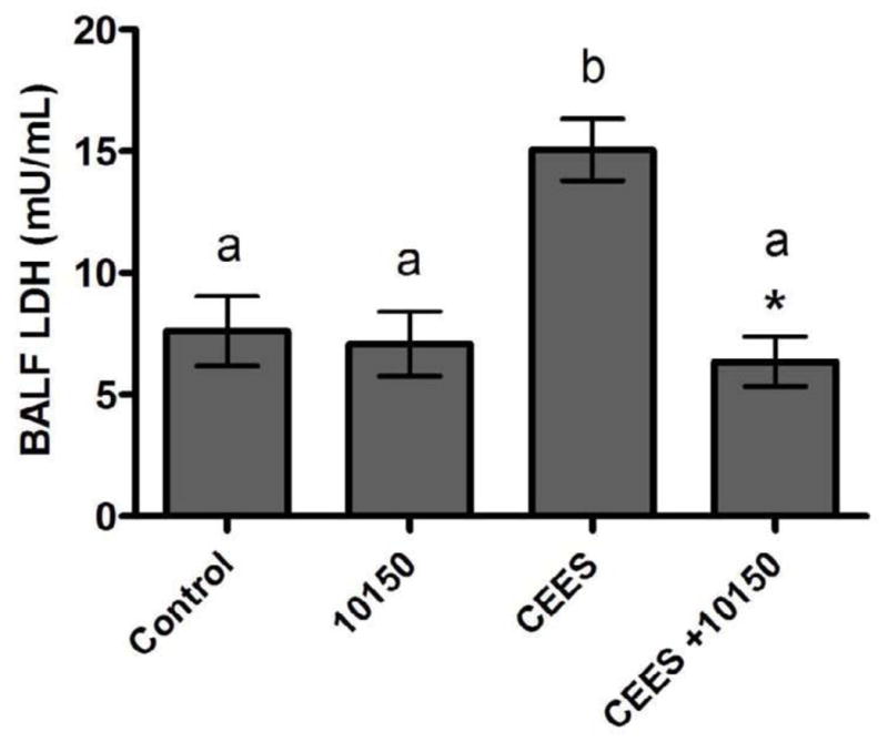
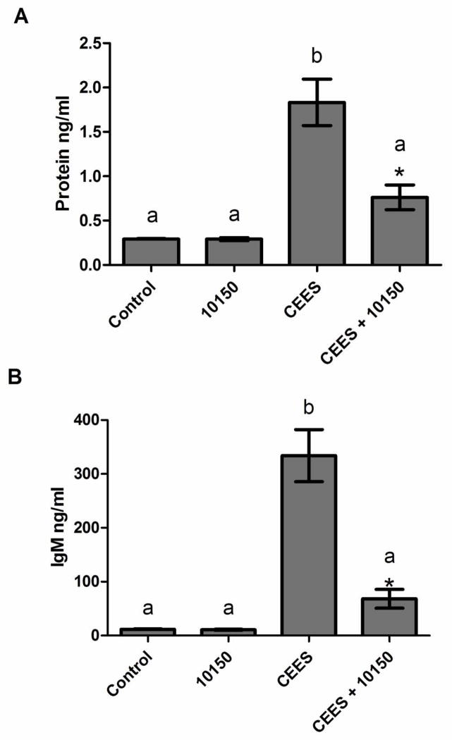
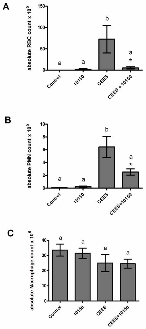
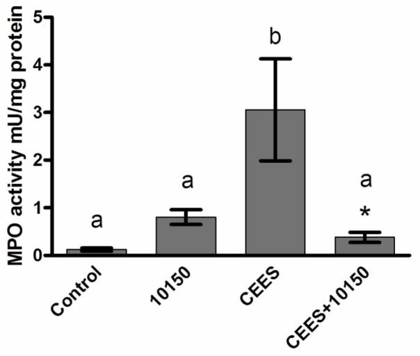
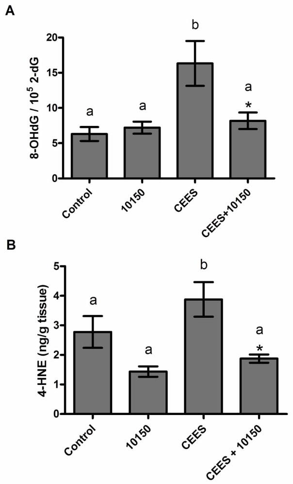
Sulfur mustard (bis-2-(chloroethyl) sulfide; SM) is a highly reactive vesicating and alkylating chemical warfare agent. A SM analog, 2-chloroethyl ethyl sulfide (CEES), has been utilized to elucidate mechanisms of toxicity and as a screen for therapeutics. Previous studies with SM and CEES have demonstrated a role for oxidative stress as well as decreased injury with antioxidant treatment. We tested whether posttreatment with the metalloporphyrin catalytic antioxidant AEOL 10150 would improve outcome in CEES-induced lung injury. Anesthetized rats inhaled 5% CEES for 15 min via a nose-only inhalation system. At 1 and 9 h after CEES exposure, rats were given AEOL 10150 (5 mg/kg, sc). At 18 h post-CEES exposure BALF lactate dehydrogenase activity, protein, IgM, red blood cells, and neutrophils were elevated but were decreased by AEOL 10150 treatment. Lung myeloperoxidase activity was increased after CEES inhalation and was ameliorated by AEOL 10150. The lung oxidative stress markers 8-OHdG and 4-HNE were elevated after CEES exposure and significantly decreased by AEOL 10150 treatment. These findings demonstrate that CEES inhalation increased lung injury, inflammation, and oxidative stress, and AEOL 10150 was an effective rescue agent. Further investigation utilizing catalytic antioxidants as treatment for SM inhalation injury is warranted.


Figures

Figure 1
Figure 1
Effect of treatment with AEOL 10150 on CEES-induced BALF LDH activity. Rats were either exposed to EtOH or 5% CEES for 15 minutes and then given PBS (1 ml/kg, SC) or AEOL 10150 (5 mg/kg, SC) treatment 1 and 9 hours after inhalation exposure. BALF was harvested 18 hours after exposure and lung injury was assessed by measuring changes in LDH levels. Columns with different letters are significantly different from one another. Asterisks denote a significant difference between CEES inhalation and CEES inhalation with AEOL 10150 treatment (p< 0.05). Data are shown as mean ± S.E.M., n=5 (control, 10150) and n=9 (CEES, CEES+10150).
Figure 2
Figure 2
Effect of AEOL 10150 on CEES-induced increases in markers of acute lung edema. Rats were either exposed to EtOH or 5% CEES for 15 minutes and then given either PBS (1 ml/kg, SC) or AEOL 10150 (5 mg/kg, SC) at 1 and 9 hours after CEES exposure. At 18 hours post exposure, rats were lavaged and levels of BALF protein (A) and IgM (B) were determined by a BCA assay and ELISA assay, respectively. Columns with different letters are significantly different from one another. Asterisks denote a significant difference between CEES inhalation and CEES inhalation with AEOL 10150 treatment (p< 0.05). Data are shown as mean ± S.E.M., protein n=6 (control, 10150) and n=16 (CEES, CEES+10150), IgM n=6 for all groups.
Figure 3
Figure 3
Effect of AEOL 10150 on CEES-induced increases in numbers of hemorrhage and inflammation. BALF was centrifuged, and cells were resuspended in PBS. Rats were either exposed to EtOH or 5% CEES for 15 minutes and then given PBS (1 ml/kg, SC) or AEOL 10150 (5 mg/kg, SC) at 1 and 9 hours after CEES exposure. At 18 hours after CEES exposure, rats were lavaged and BALF was centrifuged with resulting cell pellets resuspended in PBS. (A) Absolute numbers of BALF RBC, (B) absolute numbers of BALF PMN, and (C) absolute numbers of BALF macrophages are shown. Columns with different letters are significantly different from one another. Asterisks denote a significant difference between CEES inhalation and CEES inhalation with AEOL 10150 treatment (p< 0.05). Data are mean ± S.E.M., n=6 (control, 10150) and n=13 (CEES, CEES+10150).
Figure 4
Figure 4
Effect of AEOL 10150 on CEES-induced increases in lung tissue MPO activity. Rats were either exposed to EtOH or 5% CEES for 15 minutes and then given PBS (1 ml/kg, SC) or AEOL 10150 (5 mg/kg, SC) at 1 and 9 hours after CEES exposure. Snap-frozen lung tissue was homogenized and MPO activity measured. Columns with different letters are significantly different from one another. An asterisk denote a significant difference between CEES inhalation and CEES inhalation with AEOL 10150 treatment (p< 0.05). Data are shown as mean ± S.E.M., n=6 for all groups.
Figure 5
Figure 5
The effect of AEOL 10150 on CEES-induced lung oxidative stress. Rats were either exposed to EtOH or 5% CEES for 15 minutes and then given PBS (1 ml/kg, SC) or AEOL 10150 (5 mg/kg, SC) at 1 and 9 hours after CEES exposure. (A) Lung 2-dG and 8-OHdG levels were measured using HPLC with UV and electrochemical detection, respectively. Standard curves of 8-OHdG and 2-dG were used to determine concentrations. Data was expressed as the ratio of 8-OHdG/105 2-dG. (B) 4-HNE was measured using GC/MS analysis. Concentration of 4-HNE was determined using a standard curve generated by graphing the area ratio of 4-HNE to d3-4-HNE. An asterisk denote a significant difference between CEES inhalation and CEES inhalation with AEOL 10150 treatment (p< 0.05). 8-OHdG data were shown as mean ± S.E.M., n=12. 4-HNE data presented as mean ± S.E.M., n=11 (control and CEES), n= 5 for (10150 and CEES+10150).
Figure 6
Figure 6
Representative light photomicrographs of the lung following EtOH or CEES inhalation and subsequent PBS or AEOL 10150 treatment. Tissues were stained with hematoxylin & eosin; low magnification (100× total) views with inset at 400× total magnification are shown. Lungs from (A) control (ethanol-exposed) and (B) AEOL10150-treated rat lungs demonstrate normal airways following ethanol inhalation. (C) Shows severe damage, (D) shows moderate damage, and (E) shows minimal damage following CEES inhalation. (F) shows severe damage, (G) shows moderate damage and (H) shows minimal damage following CEES inhalation with AEOL 10150 treatment.
Figure 7
Figure 7
Plasma concentrations of AEOL10150 following a single SC injection at 5 mg/mg. A total of 15 rats were injected with AEOL 10150 and 3 rats per time point were sacrificed at 1,2,4,6, and 8 hour time points to measure AEOL 10150 levels.

Similar articles

Cited by

References

    1. Kehe K, Szinicz L. Medical aspects of sulphur mustard poisoning. Toxicology. 2005;214:198–209. - PubMed
    1. Dacre JC, Goldman M. Toxicology and pharmacology of the chemical warfare agent sulfur mustard. Pharmacol Rev. 1996;48:289–326. - PubMed
    1. Papirmeister B, Gross CL, Meier HL, Petrali JP, Johnson JB. Molecular basis for mustard-induced vesication. Fundam Appl Toxicol. 1985;5:S134–149. - PubMed
    1. Elsayed NM, Omaye ST, Klain GJ, Korte DW., Jr Free radical-mediated lung response to the monofunctional sulfur mustard butyl 2-chloroethyl sulfide after subcutaneous injection. Toxicology. 1992;72:153–165. - PubMed
    1. Elsayed NM, Omaye ST. Biochemical changes in mouse lung after subcutaneous injection of the sulfur mustard 2-chloroethyl 4-chlorobutyl sulfide. Toxicology. 2004;199:195–206. - PubMed

MeSH terms