Interaction between cartilage oligomeric matrix protein and extracellular matrix protein 1 mediates endochondral bone growth
- PMID: 20138147
- PMCID: PMC2862898
- DOI: 10.1016/j.matbio.2010.01.007
Interaction between cartilage oligomeric matrix protein and extracellular matrix protein 1 mediates endochondral bone growth
Abstract
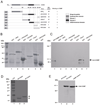
In an effort to define the biological functions of COMP, a functional genetic screen was performed. This led to the identification of extracellular matrix protein 1 (ECM1) as a novel COMP-associated partner. COMP directly binds to ECM1 both in vitro and in vivo. The EGF domain of COMP and the C-terminus of ECM1 mediate the interaction between them. COMP and ECM1 colocalize in the growth plates invivo. ECM1 inhibits chondrocyte hypertrophy, matrix mineralization, and endochondral bone formation, and COMP overcomes the inhibition by ECM1. In addition, COMP-mediated neutralization of ECM1 inhibition depends on their interaction, since COMP largely fails to overcome the ECM1 inhibition in the presence of the EGF domain of COMP, which disturbs the association of COMP and ECM1. These findings provide the first evidence linking the association of COMP and ECM1 and the biological significance underlying the interaction between them in regulating endochondral bone growth.
Copyright 2010 International Society of Matrix Biology. Published by Elsevier B.V. All rights reserved.
Figures


Similar articles
-
Cartilage oligomeric matrix protein associates with granulin-epithelin precursor (GEP) and potentiates GEP-stimulated chondrocyte proliferation.J Biol Chem. 2007 Apr 13;282(15):11347-55. doi: 10.1074/jbc.M608744200. Epub 2007 Feb 15. J Biol Chem. 2007. PMID: 17307734
-
Genetic mouse models for the functional analysis of the perifibrillar components collagen IX, COMP and matrilin-3: Implications for growth cartilage differentiation and endochondral ossification.Histol Histopathol. 2009 Aug;24(8):1067-79. doi: 10.14670/HH-24.1067. Histol Histopathol. 2009. PMID: 19554514 Review.
-
Chop (Ddit3) is essential for D469del-COMP retention and cell death in chondrocytes in an inducible transgenic mouse model of pseudoachondroplasia.Am J Pathol. 2012 Feb;180(2):727-37. doi: 10.1016/j.ajpath.2011.10.035. Epub 2011 Dec 7. Am J Pathol. 2012. PMID: 22154935 Free PMC article.
-
Extracellular matrix protein 1, a direct targeting molecule of parathyroid hormone-related peptide, negatively regulates chondrogenesis and endochondral ossification via associating with progranulin growth factor.FASEB J. 2016 Aug;30(8):2741-54. doi: 10.1096/fj.201600261R. Epub 2016 Apr 13. FASEB J. 2016. PMID: 27075243 Free PMC article.
-
Cartilage Oligomeric Matrix Protein, Diseases, and Therapeutic Opportunities.Int J Mol Sci. 2022 Aug 17;23(16):9253. doi: 10.3390/ijms23169253. Int J Mol Sci. 2022. PMID: 36012514 Free PMC article. Review.
Cited by
-
Enhanced COMP catabolism detected in serum of patients with arthritis and animal disease models through a novel capture ELISA.Osteoarthritis Cartilage. 2012 Aug;20(8):854-62. doi: 10.1016/j.joca.2012.05.003. Epub 2012 May 14. Osteoarthritis Cartilage. 2012. PMID: 22595227 Free PMC article.
-
ROS-induced PADI2 downregulation accelerates cellular senescence via the stimulation of SASP production and NFκB activation.Cell Mol Life Sci. 2022 Feb 26;79(3):155. doi: 10.1007/s00018-022-04186-5. Cell Mol Life Sci. 2022. PMID: 35218410 Free PMC article.
-
Stromal protein Ecm1 regulates ureteric bud patterning and branching.PLoS One. 2013 Dec 31;8(12):e84155. doi: 10.1371/journal.pone.0084155. eCollection 2013. PLoS One. 2013. PMID: 24391906 Free PMC article.
-
The molecular mechanism study of COMP involved in the articular cartilage damage of Kashin-Beck disease.Bone Joint Res. 2020 Sep 20;9(9):578-586. doi: 10.1302/2046-3758.99.BJR-2019-0247.R1. eCollection 2020 Sep. Bone Joint Res. 2020. PMID: 33005397 Free PMC article.
-
Endothelial Cell-Derived Lactate Triggers Bone Mesenchymal Stem Cell Histone Lactylation to Attenuate Osteoporosis.Adv Sci (Weinh). 2023 Nov;10(31):e2301300. doi: 10.1002/advs.202301300. Epub 2023 Sep 26. Adv Sci (Weinh). 2023. PMID: 37752768 Free PMC article.
References
-
- Bhalerao J, Tylzanowski P, Filie JD, Kozak CA, Merregaert J. Molecular cloning, characterization, and genetic mapping of the cDNA coding for a novel secretory protein of mouse. Demonstration of alternative splicing in skin and cartilage. J Biol Chem. 1995;270:16385–16394. - PubMed
-
- Briggs MD, Hoffman SM, King LM, Olsen AS, Mohrenweiser H, Leroy JG, Mortier GR, Rimoin DL, Lachman RS, Gaines ES. Pseudoachondroplasia and multiple epiphyseal dysplasia due to mutations in the cartilage oligomeric matrix protein gene. Nature. 1995;10:330–336. - PubMed
Publication types
MeSH terms
Substances
Grants and funding
LinkOut - more resources
Full Text Sources
Molecular Biology Databases
Research Materials
Miscellaneous

