. 2010 May;31(13):3736-43.
doi: 10.1016/j.biomaterials.2010.01.058. Epub 2010 Feb 6.

Bioactive hydrogels made from step-growth derived PEG-peptide macromers

Affiliations

Bioactive hydrogels made from step-growth derived PEG-peptide macromers

Jordan S Miller et al. Biomaterials. 2010 May.

Abstract

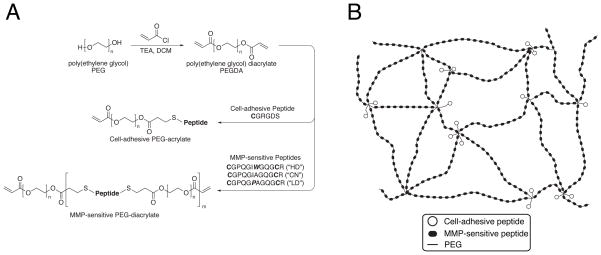
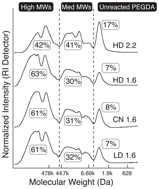
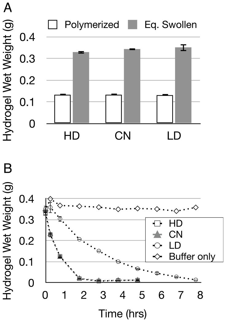
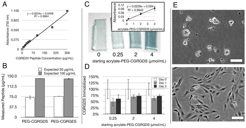
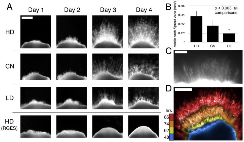
Synthetic hydrogels based on poly(ethylene glycol) (PEG) have been used as biomaterials for cell biology and tissue engineering investigations. Bioactive PEG-based gels have largely relied on heterobifunctional or multi-arm PEG precursors that can be difficult to synthesize and characterize or expensive to obtain. Here, we report an alternative strategy, which instead uses inexpensive and readily available PEG precursors to simplify reactant sourcing. This new approach provides a robust system in which to probe cellular interactions with the microenvironment. We used the step-growth polymerization of PEG diacrylate (PEGDA, 3400Da) with bis-cysteine matrix metalloproteinase (MMP)-sensitive peptides via Michael-type addition to form biodegradable photoactive macromers of the form acrylate-PEG-(peptide-PEG)(m)-acrylate. The molecular weight (MW) of these macromers is controlled by the stoichiometry of the reaction, with a high proportion of resultant macromer species greater than 500kDa. In addition, the polydispersity of these materials was nearly identical for three different MMP-sensitive peptide sequences subjected to the same reaction conditions. When photopolymerized into hydrogels, these high MW materials exhibit increased swelling and sensitivity to collagenase-mediated degradation as compared to previously published PEG hydrogel systems. Cell-adhesive acrylate-PEG-CGRGDS was synthesized similarly and its immobilization and stability in solid hydrogels was characterized with a modified Lowry assay. To illustrate the functional utility of this approach in a biological setting, we applied this system to develop materials that promote angiogenesis in an ex vivo aortic arch explant assay. We demonstrate the formation and invasion of new sprouts mediated by endothelial cells into the hydrogels from embedded embryonic chick aortic arches. Furthermore, we show that this capillary sprouting and three-dimensional migration of endothelial cells can be tuned by engineering the MMP-susceptibility of the hydrogels and the presence of functional immobilized adhesive ligands (CGRGDS vs. CGRGES peptide). The facile chemistry described and significant cellular responses observed suggest the usefulness of these materials in a variety of in vitro and ex vivo biologic investigations, and may aid in the design or refinement of material systems for a range of tissue engineering approaches.


Figures

Figure 1
Figure 1
(a) Synthetic scheme. PEG 3400 is reacted with acryloyl chloride to form PEGDA, which is then reacted with cysteine-bearing peptides via Michael-type addition to form cell-adhesive or, in a separate reaction, MMP-sensitive PEG-acrylate macromers. Reaction stoichiometry controls the molecular weight and polydispersity of the resultant species during step-growth polymerization. (b) Schematic illustration of hydrogel structure. Photopolymerization of the photoactive precursors from (a) yields bioactive hydrogels with multiple MMP-sensitive peptides per backbone chain, with pendant cell-adhesive ligands tethered from sites of acrylate crosslinking.
Figure 2
Figure 2
GPC analysis of MMP-sensitive PEG-diacrylates plotted against PEG MW standards. Highly degradable (“HD”) peptide reacted with a 2.2 molar excess of PEGDA (uppermost curve) via step-growth polymerization resulted in more than 80% conjugation (sum of Medium and High MWs). Reaction of MMP-sensitive peptides with a 1.6 molar excess of PEGDA (remaining three curves) resulted in more than 90% conjugation, with a majority of the molecular weight species greater than 500 kDa. When subjected to the same reaction stoichiometry, HD, collagen native (“CN”), and least degradable (“LD”) PEG-peptide conjugates show nearly identical polydispersity.
Figure 3
Figure 3
(a) MMP-sensitive hydrogels (made from HD, CN, or LD peptides) were polymerized at 10% w/w and then swollen to equilibrium over 36 hours (n=3, Eq. Swollen in figure). Bars indicate standard deviation. (b) Swollen hydrogels were degraded in 0.2 mg/mL collagenase (n=3) or incubated in buffer (n=1) up to 8 hours while their wet weight was monitored. Note that HD and CN have overlapping degradation curves. Bars indicate standard deviation.
Figure 4
Figure 4
The immobilization efficiency and stability of acrylate-PEG-peptide macromers in PEGDA gels was assessed with a modified Lowry Assay for total protein concentration, as well as HUVEC seeding. (a) The Lowry assay, typically only used for large proteins, produced a linear standard curve from the short, soluble CGREDV peptide, even at low concentrations. (b) This standard curve was used to quantify the solution-based concentration of acrylate-PEG-CGRGDS and acrylate-PEG-CGRGES macromers, with a deviation from expected of 40–50%, with values comparable between both peptides. Bars indicate standard error. (c) gross appearance of hydrogel slabs after modified Lowry Assay in situ showing characteristic blue color with starting peptide concentration (μmol/mL). The linear dependence on concentration was also valid in solid hydrogels (inset, bars indicate standard deviation). (d) The assay tracked CGRGDS retention over time within hydrogels. A large percent of RGDS was lost on the first day during hydrogel equilibrium swelling. The remaining peptide was stable for at least 2 more days in the gel (n=3 for all samples), with up to 75% retention. Bars indicate standard deviation. (e) HUVEC morphology on PEGDA hydrogels with 4.0 μmol/mL PEG-CGRGES (top) or PEG-CGRGDS (bottom) 24 hours post-seeding. Scale Bars = 25 μm.
Figure 5
Figure 5
(a) Representative images of chick aortic arch ring explants sprouting into hydrogels over time. In 8-wt% gels with 1.0 μmol/mL CGRGDS density, angiogenic sprouting varies with the MMP-susceptibility of the hydrogel backbone. No detectable sprouting occurred in negative control hydrogels containing RGES instead of RGDS peptide. Scale bar for all images = 250 μm. (b) Quantification of sprout area at Day 4, n=6 per condition. Mean with standard deviation, all comparisons are significant, p < 0.003 by one-way ANOVA and Tukey’s HSD post-hoc testing. (c) Fluorescent staining with lectin-rhodamine implicates endothelial cells as a principal component of the angiogenic sprouts in these hydrogels. Scale Bar = 100 μm. (d) Composite image of selected frames during sprouting time-course by dark field imaging (see supplemental Movie 1), false colored then overlaid here to aid in time visualization. Blue, yellow, orange, red = 48, 62, 74, 86 hours respectively. Scale Bar = 250 μm.

References

    1. Lutolf MP, Hubbell JA. Synthetic biomaterials as instructive extracellular microenvironments for morphogenesis in tissue engineering. Nat Biotechnol. 2005;23:47–55. - PubMed
    1. Tibbitt MW, Anseth KS. Hydrogels as extracellular matrix mimics for 3D cell culture. Biotechnol Bioeng. 2009;103:655–63. - PMC - PubMed
    1. Hill-West JL, Chowdhury SM, Sawhney AS, Pathak CP, Dunn RC, Hubbell JA. Prevention of postoperative adhesions in the rat by in situ photopolymerization of bioresorbable hydrogel barriers. Obstet Gynecol. 1994;83:59–64. - PubMed
    1. West J, Hubbell J. Polymeric biomaterials with degradation sites for proteases involved in cell migration. Macromolecules. 1999;32:241–4.
    1. Lutolf MP, Hubbell JA. Synthesis and physicochemical characterization of end-linked poly(ethylene glycol)-co-peptide hydrogels formed by Michael-type addition. Biomacromolecules. 2003;4:713–22. - PubMed

Publication types