Review
. 2010 Apr;42(4):261-71.
doi: 10.1016/j.dld.2010.01.006. Epub 2010 Feb 6.

Polycystic liver diseases

Affiliations
Review

Polycystic liver diseases

P Onori et al. Dig Liver Dis. 2010 Apr.

Abstract

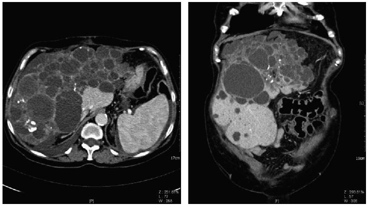
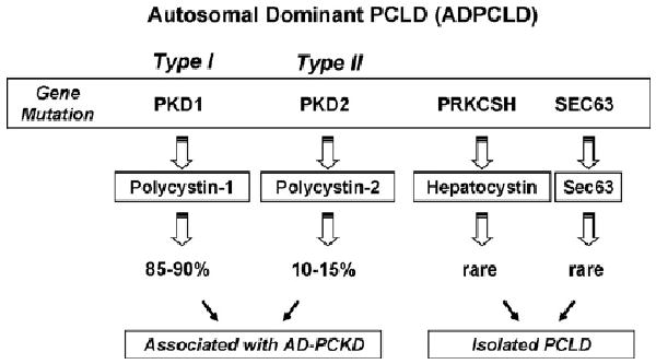
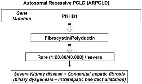
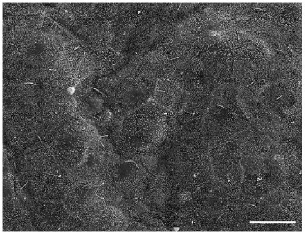
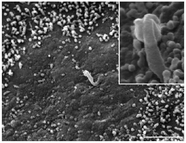
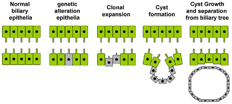
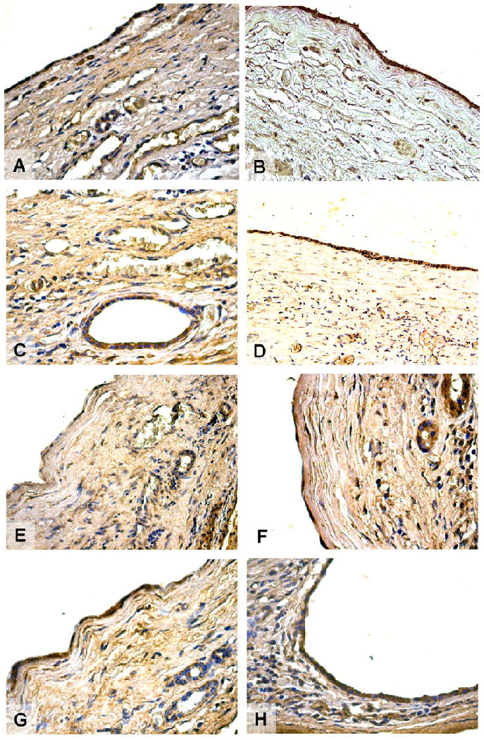
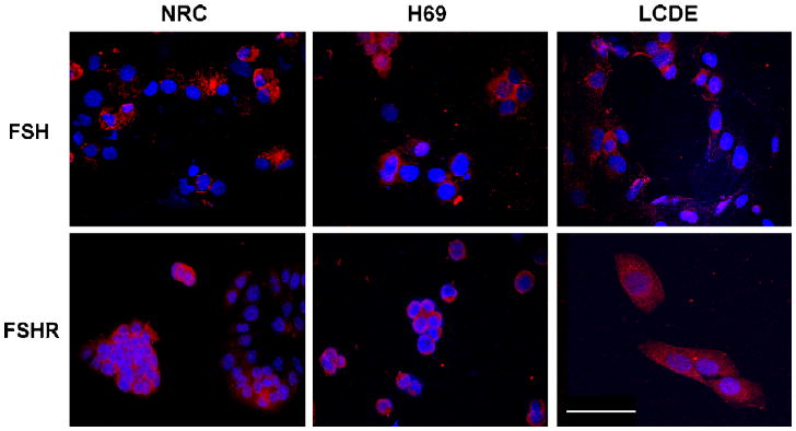
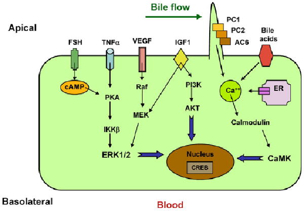
Polycystic liver diseases (PCLDs) are genetic disorders with heterogeneous etiologies and a range of phenotypic presentations. PCLD exhibits both autosomal or recessive dominant pattern of inheritance and is characterized by the progressive development of multiple cysts, isolated or associated with polycystic kidney disease, that appear more extensive in women. Cholangiocytes have primary cilia, functionally important organelles (act as mechanosensors) that are involved in both normal developmental and pathological processes. The absence of polycystin-1, 2, and fibrocystin/polyductin, normally localized to primary cilia, represent a potential mechanism leading to cyst formation, associated with increased cell proliferation and apoptosis, enhanced fluid secretion, abnormal cell-matrix interactions, and alterations in cell polarity. Proliferative and secretive activities of cystic epithelium can be regulated by estrogens either directly or by synergizing growth factors including nerve growth factor, IGF1, FSH and VEGF. The abnormalities of primary cilia and the sensitivity to proliferative effects of estrogens and different growth factors in PCLD cystic epithelium provide the morpho-functional basis for future treatment targets, based on the possible modulation of the formation and progression of hepatic cysts.


Conflict of interest statement

Conflict of interest statement: All the authors declare that there are no conflicts of interest.

Figures

Fig. 1
Fig. 1
Abdominal computed tomography (axial and frontal orientation respectively) of an adult with ADPCLD showing extensive cyst formation in liver parenchyma (images kindly provided by Prof. C. Catalano and Dr. V. Cardinale).
Fig. 2
Fig. 2
Diagram of the genetics of polycystic liver disease in its autosomic dominant form, with mutation on the genes PKD1, PKD2, PRKCSH and SEC63.
Fig. 3
Fig. 3
Diagram of the genetic of polycystic liver disease in its subtype autosomic recessive linked to modifications in the gene PKHD1 that codes for the protein fibrocystin.
Fig. 4
Fig. 4
SEM of luminal surface of a small hepatic cyst of ADPKD liver. The epithelium lining small cyst (1 cm maximum diameter) shows a carpet of regular microvilli and typical primary cilia comparable with a normal biliary epithelium. Bar 10 μm.
Fig. 5
Fig. 5
SEM of epithelial surface of a cyst with a 2–2.5 cm maximum diameter shows less dense microvilli and clear areas become visible. The cilium is absent or shorter than the typical structure and often showed alteration of the apical zone. In the box, at higher magnification a short and abnormal cilium is evident. Bar 2 μm.
Fig. 6
Fig. 6
Diagram depicting the genetic basis of cyst formation in ADPCLD. The biliary epithelium in a normal person has both alleles of PKD genes. In patients with ADPCLD, there is a germ-line mutation of one of the alleles (‘first hit’). Some cells within the duct acquire a mutation in the second allele (‘second hit’), resulting in increased proliferative activity in these cells, leading ultimately to cyst formation. The cells lining these cysts are monoclonal and are homozygous for PKD mutation.
Fig. 7
Fig. 7
Immunohistochemistry for ER-α (A), ER-β (B), IGF-1 (C), IGF1-R (D), FSH (E), FSHR (F), VEGF-A (G), and VEGF-C (H) in hepatic cysts from patients with ADPCLD. Cholangiocytes of reactive bile ducts close to the cysts are positive for both estrogen receptors. The epithelium lining small and large cysts showed strong positivity for ER-α and ER-β located at cytoplasmic levels. Both IGF1 and its receptor showed positive immunolocalisation in biliary epithelium. The expression of the hormone FSH is also present in both small and large cysts. A stronger immunolocalisation is found for FSHR in the biliary epithelium that lines hepatic cysts. Furthermore, biliary epithelium of small and large cysts shows an high positivity for VEGF-A and VEGF-C, that play an important role in the growth of cysts and in the progression of PCLD. Original magnification 20 ×.
Fig. 8
Fig. 8
Immunofluorescence in immortalized cell cultures from normal rat cholangiocytes (NRC), normal human cholangiocytes (H69) and cholangiocytes from hepatic cysts (LCDE) shows the presence of positivity for FSH and FSHR. Original magnification 40×. Bar 200 μm.
Fig. 9
Fig. 9
Diagram depicting putative pathways in polycystic liver disease. Dysregulation of [Ca2+]i, increased concentrations of cAMP and upregulation of FSH, IGF1, VEGF, and TNFα occur in cells bearing PKD mutations. Increased accumulation of cAMP in polycystic livers may result from disruption of the polycystin complex, as PC1 may act as a Gi protein-coupled receptor and stimulation of Ca2+ inhibitable AC6. Increased cAMP levels contribute to cystogenesis by stimulating chloride and fluid secretion. In addition, cAMP stimulates mitogen-activated protein kinase/extracellular regulated kinase (MAPK/ERK) signalling in cyst-derived cells.

Similar articles

Cited by

References

    1. Everson GT, Helmke SM, Doctor B. Advances in management of polycystic liver disease. Expert Rev Gastroenterol Hepatol. 2008;2:563–76. - PubMed
    1. Masyuk T, LaRusso N. Polycystic liver disease: new insights into disease pathogenesis. Hepatology. 2006;43:906–8. - PubMed
    1. Everson GT, Taylor MR, Doctor RB. Polycystic disease of the liver. Hepatology. 2004;40:774–82. - PubMed
    1. Torres VE, Harris PC, Pirson Y. Autosomal dominant polycystic kidney disease. Lancet. 2007;369:1287–301. - PubMed
    1. Xu C, Rossetti S, Jiang L, et al. Human ADPKD primary cyst epithelial cells with a novel, single codon deletion in the PKD1 gene exhibit defective ciliary polycystin localization and loss of flow-induced Ca2+ signaling. Am J Physiol Renal Physiol. 2007;292:F930–45. - PMC - PubMed

Publication types

Substances