. 2010 Apr 6:1323:18-32.
doi: 10.1016/j.brainres.2010.01.081. Epub 2010 Feb 6.

Cocoa-enriched diets enhance expression of phosphatases and decrease expression of inflammatory molecules in trigeminal ganglion neurons

Affiliations

Cocoa-enriched diets enhance expression of phosphatases and decrease expression of inflammatory molecules in trigeminal ganglion neurons

Ryan J Cady et al. Brain Res. .

Abstract

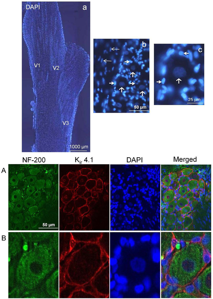
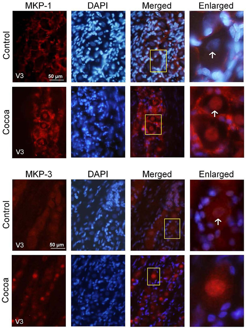
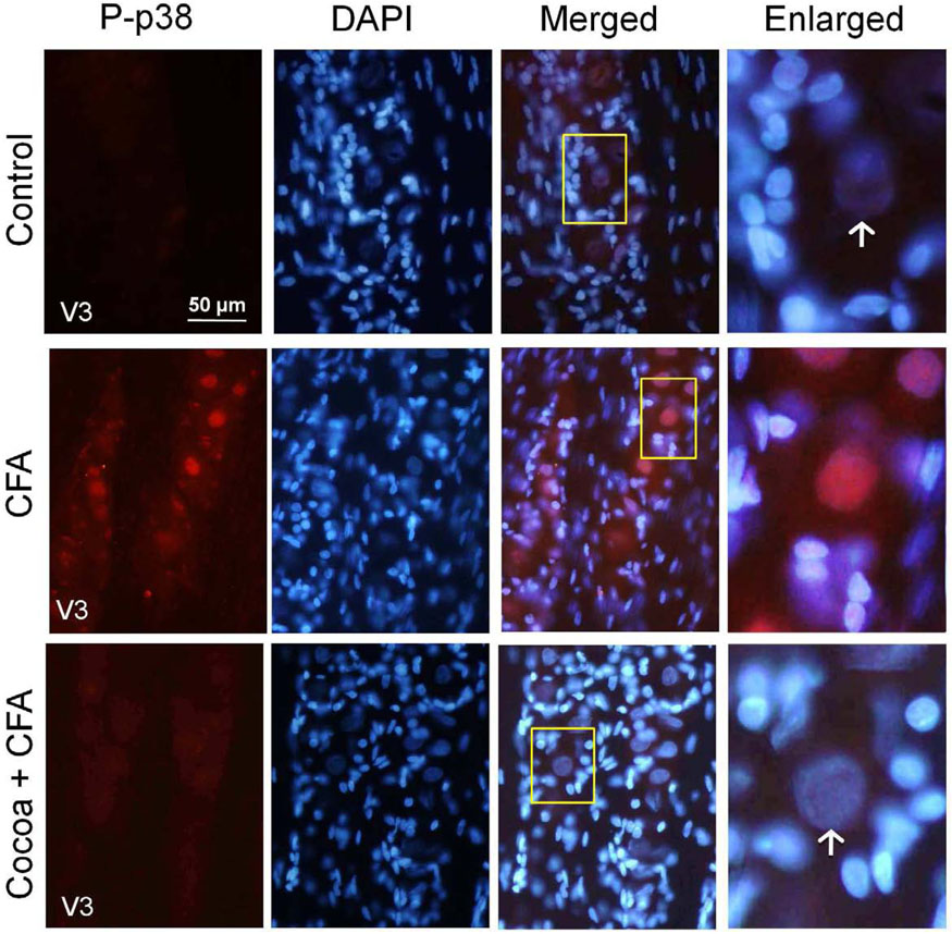
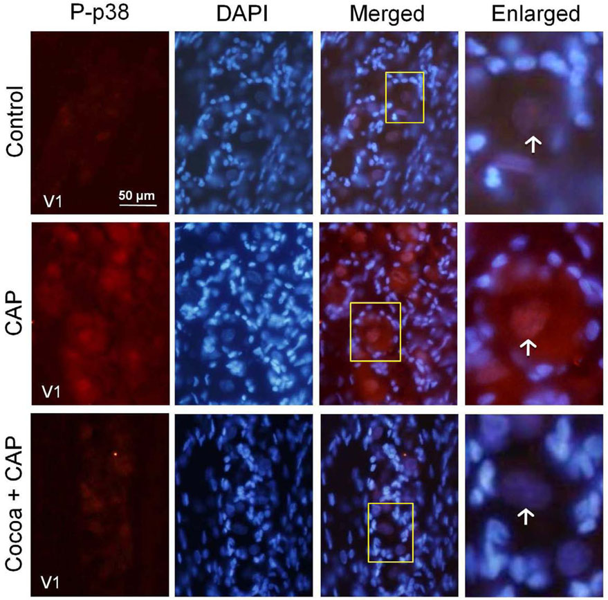
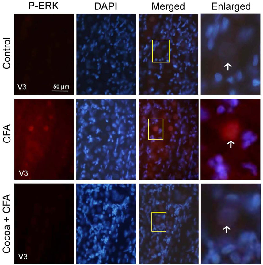
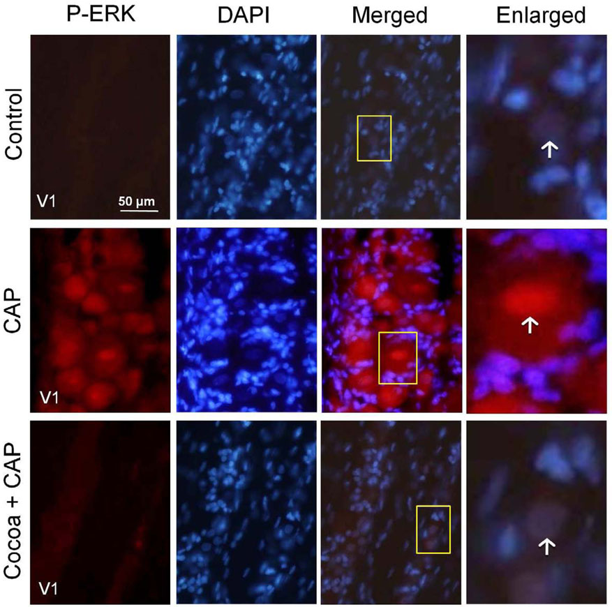
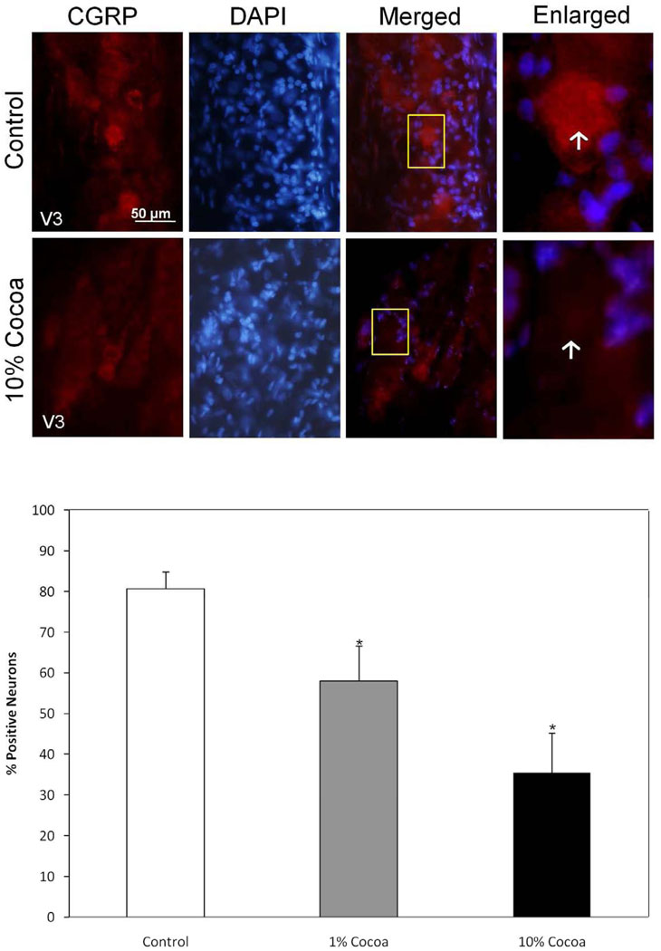
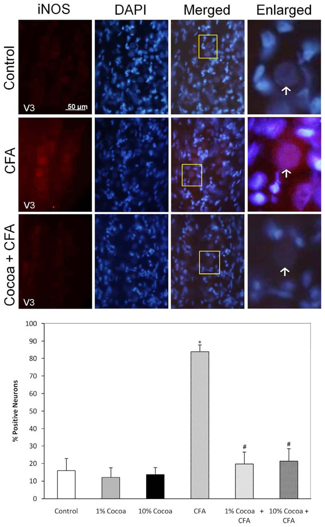
Activation of trigeminal nerves and release of neuropeptides that promote inflammation are implicated in the underlying pathology of migraine and temporomandibular joint (TMJ) disorders. The overall response of trigeminal nerves to peripheral inflammatory stimuli involves a balance between enzymes that promote inflammation, kinases, and those that restore homeostasis, phosphatases. The goal of this study was to determine the effects of a cocoa-enriched diet on the expression of key inflammatory proteins in trigeminal ganglion neurons under basal and inflammatory conditions. Rats were fed a control diet or an isocaloric diet enriched in cocoa for 14days prior to an injection of noxious stimuli to cause acute or chronic excitation of trigeminal neurons. In animals fed a cocoa-enriched diet, basal levels of the mitogen-activated kinase (MAP) phosphatases MKP-1 and MKP-3 were elevated in neurons. Importantly, the stimulatory effects of acute or chronic peripheral inflammation on neuronal expression of the MAPK p38 and extracellular signal-regulated kinases (ERK) were significantly repressed in response to cocoa. Similarly, dietary cocoa significantly suppressed basal neuronal expression of calcitonin gene-related peptide (CGRP) as well as stimulated levels of the inducible form of nitric oxide synthase (iNOS), proteins implicated in the underlying pathology of migraine and TMJ disorders. To our knowledge, this is the first evidence that a dietary supplement can cause upregulation of MKP, and that cocoa can prevent inflammatory responses in trigeminal ganglion neurons. Furthermore, our data provide evidence that cocoa contains biologically active compounds that would be beneficial in the treatment of migraine and TMJ disorders.


Figures

FIG. 1
FIG. 1
Organization and identification of neurons and glia in the trigeminal ganglion. The cellular organization of the trigeminal ganglion is shown in the top panels. (a) A longitudinal section of a trigeminal ganglion stained with the fluorescent dye DAPI to identify cell nuclei is shown at 40× magnification. The relative locations of the V1, V2, and V3 regions are indicated. (b) The cell bodies of neuronal cells are organized into bands or clusters within the ganglion. A band of neuronal (vertical arrows) and satellite glial cells (thick horizontal arrows) is shown at 400× magnification. Also visible are Schwann cells (thin horizontal arrows). (c) An enlarged image of a functional unit consisting of a single neuron (vertical arrow) surrounded by several satellite glia cells (horizontal arrows) is shown. Bottom panels: A 400× magnification of a neuron-satellite glia enriched region costained with antibodies against the neuronal cell marker NF-200 and the satellite glial cell marker Kir 4.1, as well as DAPI is shown (A). An enlarged image of a functional unit is shown in panel B. A merged image of the same sections stained for NF-200, Kir 4.1, and DAPI staining is shown in the bottom right panels.
FIG. 2
FIG. 2
MPK-1 and MKP-3 expression are increased in rats fed cocoa enriched diets. A section of the posterolateral portion of the ganglia (V3) was obtained from untreated animals (Control) or animals fed 10% cocoa (g/g) for 14 days. A 400× magnification of neuron-satellite glia enriched regions stained for MKP-1 (top panel) or MKP-3 (bottom panel) are shown in the left panels. The next three panels represent the same section stained with the nuclear dye DAPI, a merged image of MKP-1 or MKP-3 and DAPI staining, and an enlarged image from the tissue outlined by the yellow box on the merged image (right panel). Neurons are identified by vertical arrows.
FIG. 3
FIG. 3
P-p38 expression in response to CFA is decreased in rats fed cocoa enriched diets. Sections from the posterolateral portion (V3) of the ganglion were obtained from untreated animals (Control), animals injected with CFA for five days, or animals fed 10% cocoa for 14 days and injected with CFA for 5 days prior to harvesting the ganglia (Cocoa + CFA). A 400× magnification of neuron-satellite glia enriched regions stained for P-p38 is shown in the left panels. The next three panels represent the same section stained with the nuclear dye DAPI, a merged image showing both P-p38 and DAPI staining, and an enlarged image taken from the tissue outlined by the yellow box on the merged image (right panel). Neurons are identified by vertical arrows.
FIG. 4
FIG. 4
Capsaicin stimulation of P-p38 expression is decreased in rats fed cocoa enriched diets. Sections of the anterolateral portion (V1) of the ganglia were obtained from untreated animals (Control), animals injected with capsaicin (CAP) for two hours, or animals fed 10% cocoa (g/g) diet for 14 days and injected with capsaicin for 2 hours prior to harvesting the ganglia (Cocoa + CAP). A 400× magnification of neuron-satellite glia enriched regions stained for P-p38 is shown in the left panels. The next three panels represent the same section stained with the nuclear dye DAPI, a merged image showing both P-p38 and DAPI staining, and an enlarged image taken from the tissue outlined by the yellow box on the merged image (right panel). Neurons are identified by vertical arrows.
FIG. 5
FIG. 5
The stimulatory effects of CFA on P-ERK expression are repressed in rats fed cocoa enriched diets. Sections from the posterolateral portion (V3) of the ganglion were obtained from untreated animals (Control), animals injected with CFA for five days, or animals fed 10% cocoa for 14 days and injected with CFA for 5 days prior to harvesting the ganglia (Cocoa + CFA). A 400× magnification of neuron-satellite glia enriched regions stained for P-ERK is shown in the left panels. The next three panels represent the same section stained with the nuclear dye DAPI, a merged image showing both P-ERK and DAPI staining, and an enlarged image taken from the tissue outlined by the yellow box on the merged image (right panel). Neurons are identified by vertical arrows.
FIG. 6
FIG. 6
Increases in P-ERK expression in response to capsaicin (CAP) are repressed in rats fed cocoa enriched diets. Sections of the anterolateral portion (V1) of the ganglia were obtained from untreated animals (Control), animals injected with capsaicin (CAP) for two hours, or animals fed 10% cocoa (g/g) diet for 14 days and injected with capsaicin for 2 hours prior to harvesting the ganglia (Cocoa + CAP). A 400× magnification of neuron-satellite glia enriched regions stained for P-ERK is shown in the left panels. The next three panels represent the same section stained with the nuclear dye DAPI, a merged image showing both P-ERK and DAPI staining, and an enlarged image taken from the tissue outlined by the yellow box on the merged image (right panel). Neurons are identified by vertical arrows.
FIG. 7
FIG. 7
Effect of cocoa-enriched diets on the expression of MKP and MAPK in trigeminal ganglion neurons and satellite glial cells. The data are reported as an average percentage ± SEM (n = 3 independent experiments) of the number of immunopositive neurons, which were identified by nuclear staining with DAPI. Percentage of trigeminal ganglion neurons staining positive for MKP-1 or MKP-3 (A) Percentage of trigeminal ganglion neurons staining positive for P-p38 and P-ERK under basal, unstimulated conditions or in response to cocoa, in response to CFA and cocoa, or in response to capsaicin (CAP) and cocoa (B). * p < 0.01 when compared to control while # p < 0.01 when compared to stimulation.
FIG. 8
FIG. 8
Basal CGRP expression is decreased in rats fed cocoa enriched diets. A section of the posterolateral portion of the ganglia (V3) was obtained from untreated animals (Control) or animals fed 10% cocoa (g/g) for 14 days. A 400× magnification of neuron-satellite glia enriched regions stained for CGRP is shown in the left panels. The next three panels represent the same section stained with the nuclear dye DAPI, a merged image showing both CGRP and DAPI staining, and an enlarged image taken from the tissue outlined by the yellow box on the merged image (right panel). Neurons are identified by vertical arrows. Bottom panel: Effects of cocoa-enriched diets on the expression of CGRP in trigeminal ganglion neurons. The data are reported as an average percentage ± SEM (n = 3 independent experiments) of the number of immunopositive neurons compared to the total number of neurons as identified by nuclear staining with DAPI. * p < 0.01 when compared to control.
FIG. 9
FIG. 9
Stimulated iNOS expression in response to CFA is repressed in rats fed cocoa enriched diets. A section of the posterolateral portion of the ganglia (V3) was obtained from untreated animals (Control), or animals injected with CFA for five days, or animals fed 10% cocoa for 14 days and injected with CFA for 5 days prior to harvesting the ganglia (Cocoa + CFA). A 400× magnification of neuron-satellite glia enriched regions stained for iNOS is shown in the left panels. The next three panels represent the same section stained with the nuclear dye DAPI, a merged image showing both iNOS and DAPI staining, and an enlarged image taken from the tissue outlined by the yellow box on the merged image (right panel). Neurons are identified by vertical arrows. Bottom panel: Effects of cocoa-enriched diets on the expression of iNOS in trigeminal ganglion neurons. The data are reported as an average percentage ± SEM (n = 3 independent experiments) of the number of immunopositive neurons compared to the total number of neurons as identified by nuclear staining with DAPI. * p < 0.01 when compared to control while # p < 0.01 compared to stimulation.

References

    1. Abbey MJ, Patil VV, Vause CV, Durham PL. Repression of calcitonin gene-related peptide expression in trigeminal neurons by a Theobroma cacao extract. J Ethnopharmacol. 2008;115:238–248. - PMC - PubMed
    1. Bhat N, Feinstein D, Shen Q, Bhat A. p38 MAPK-mediated transcriptional activation of inducible nitric-oxide synthase in glial cells. Roles of nuclear factors, nuclear factor kappa B, cAMP response element-binding protein, CCAAT/enhancerbinding protein-beta, and activating transcription factor-2. J Biol Chem. 2002;277:29584–29592. - PubMed
    1. Buzzi M. Trigeminal pain pathway: peripheral and central activation as experimental models of migraine. Funtional Neurology. 2001;16:77–81. - PubMed
    1. Carlsson G, LeResche L. Epidemiology of temporomandibular disorders. In: Sessle PS, Dionne RA, editors. Temporomandibular disorders and related pain conditions. Progress in pain research and management. Vol., B. Seattle: IASP Press; 1995. pp. 211–226.
    1. Caterina M, Schumacher M, Tominaga M, Rosen T, Julius D. The capsaicin receptor: a heat-activated ion channel in the pain pathway. Nature. 1997;389:816–824. - PubMed

Publication types

Substances