Inflammatory changes in the central nervous system are associated with behavioral impairment in Plasmodium berghei (strain ANKA)-infected mice
- PMID: 20138873
- PMCID: PMC3111979
- DOI: 10.1016/j.exppara.2010.02.002
Inflammatory changes in the central nervous system are associated with behavioral impairment in Plasmodium berghei (strain ANKA)-infected mice
Abstract
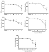
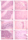
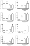
Experimental cerebral malaria is a neuroinflammatory condition that results from the host immune response to the parasite. Using intravital microscopy, we investigated leukocyte recruitment in the brain microcirculation and the temporal relationship of this process to the behavioral changes observed in Plasmodium berghei (strain ANKA)-infected C57Bl/6 mice. We found that leukocyte recruitment was increased from day 5 post-infection (p.i.) onwards. Histopathological changes and increased levels of inflammatory cytokines in the brain were also observed. Behavioral performance evaluated by the SHIRPA protocol showed functional impairment from day 6 p.i. onwards. Thus, early leukocyte migration into the brain and associated inflammatory changes may be involved in neurological impairment in parasite-infected C57Bl/6 mice.
Figures


References
-
- Bagot S, Idrissa Boubou M, Campino S, Behrschmidt C, Gorgette O, Guenet JL, Penha-Goncalves C, Mazier D, Pied S, Cazenave PA. Susceptibility to experimental cerebral malaria induced by Plasmodium berghei ANKA in inbred mouse strains recently derived from wild stock. Infection and Immunity. 2002;70:2049–2056. - PMC - PubMed
-
- Barcelos LS, Talvani A, Teixeira AS, Vieira LQ, Cassali GD, Andrade SP, Teixeira MM. Impaired inflammatory angiogenesis, but not leukocyte influx, in mice lacking TNFR1. Journal of Leukocyte Biology. 2005;78:352–358. - PubMed
-
- Belnoue E, Kayibanda M, Vigario AM, Deschemin JC, van Rooijen N, Viguier M, Snounou G, Rénia L. On the pathogenic role of brain-sequestered alphabeta CD8+ T cells in experimental cerebral malaria. Journal of Immunology. 2002;169:6369–6375. - PubMed
-
- Campanella GS, Tager AM, El Khoury JK, Thomas SY, Abrazinski TA, Manice LA, Colvin RA, Luster AD. Chemokine receptor CXCR3 and its ligands CXCL9 and CXCL10 are required for the development of murine cerebral malaria. Proceedings of the National Academy of Sciences of the United States of America. 2008;105:4814–4819. - PMC - PubMed
-
- Cutrullis RA, Postan M, Petray PB, Corral RS. Timing of expression of inflammatory mediators in skeletal muscles from mice acutely infected with the RA strain of Trypanosoma cruzi. Pathobiology. 2009;76:170–180. - PubMed
Publication types
MeSH terms
Substances
Grants and funding
LinkOut - more resources
Full Text Sources

