. 2010 Mar 19;584(6):1126-32.
doi: 10.1016/j.febslet.2010.02.003. Epub 2010 Feb 6.

A molecular mechanism for proton-dependent gating in KcsA

Affiliations

A molecular mechanism for proton-dependent gating in KcsA

Luis G Cuello et al. FEBS Lett. .

Abstract

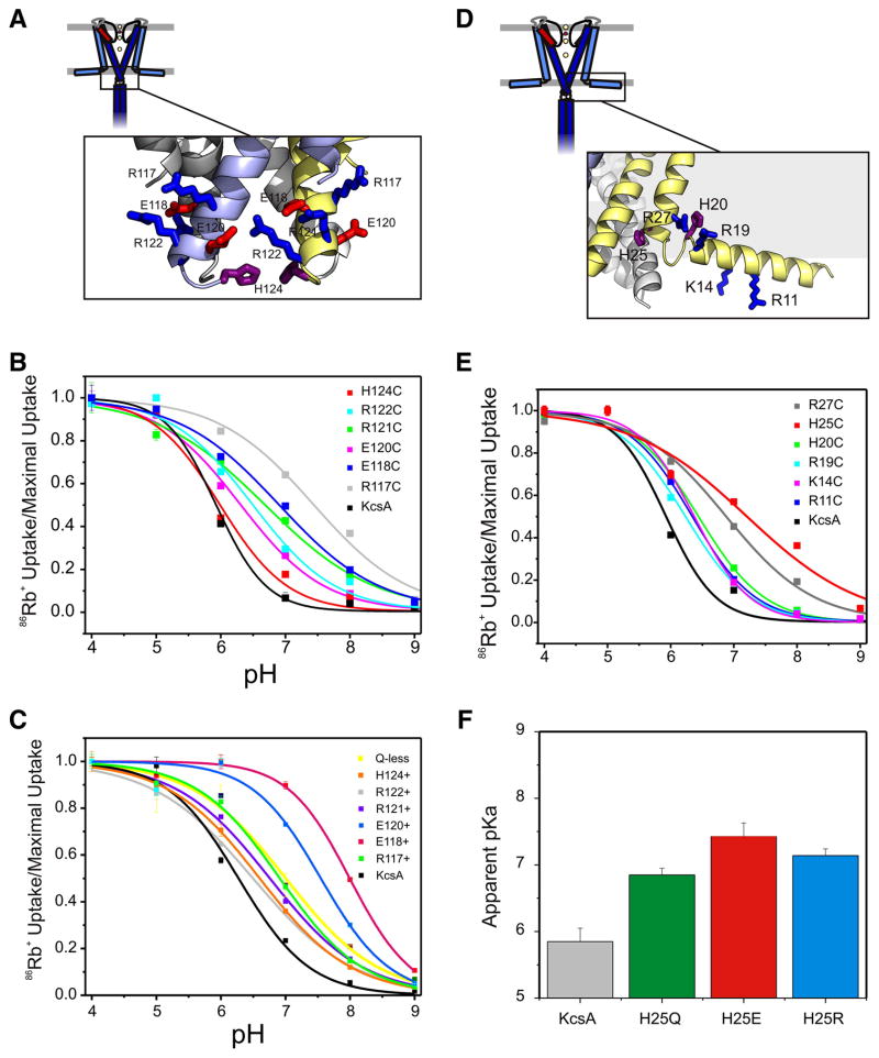
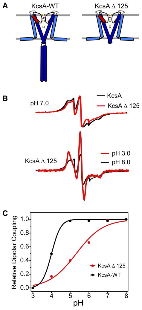
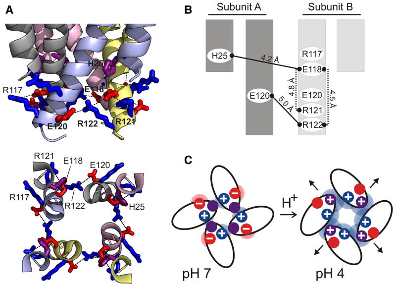
Activation gating in KcsA is elicited by changes in intracellular proton concentration. Thompson et al. identified a charge cluster around the inner gate that plays a key role in defining proton activation in KcsA. Here, through functional and spectroscopic approaches, we confirmed the role of this charge cluster and now provide a mechanism of pH-dependent gating. Channel opening is driven by a set of electrostatic interactions that include R117, E120 and E118 at the bottom of TM2 and H25 at the end of TM1. We propose that electrostatic compensation in this charge cluster stabilizes the closed conformation at neutral pH and that its disruption at low pH facilitates the transition to the open conformation by means of helix-helix repulsion.


Figures

Figure 1
Figure 1. A charge cluster near the inner bundle gate is part of the channel pH sensor
A, D. Ribbon representation of the intracellular bundle region of KcsA, ionizable residues are shown in licorice representation with basic side chains in blue, acidic side chains in red and histidines in purple. B. 86Rb uptake experiments for individual neutralization mutants in the TM2 cluster. Each pH-dependent uptake experiment was fitted to a Hill equation and activation pKas (EC50) were calculated. Activation pKa: WT=5.89±0.2, R117C=7.46±0.2, E118C=6.83±0.1, E120C=6.2±0.1, R121C=6.47±0.2, R122C=6.41±0.2, H124C=5.89±0.2. C. 86Rb uptake experiments of individual re-introduced charges in a charge-less background mutant lacking the C-terminal domain. Uptake experiments were fitted to a Hill equation and activation pKa were calculated: KcsA=6.18±0.1, R117+=6.94±0.2, E118+=7.98±0.1, E120+=7.69±0.2, R121+=6,74±0.2, R122+=6.39±0.2, H124+=6.56±0.1 and Q-less=6.7±0.2. E. pH-dependent 86Rb uptake experiments of individual neutralization mutants of TM1 N-terminus and amphipatic helix. Data were fitted to a Hill equation and activation pKa were calculated. Activation pKa: Wt=5.69±0.2, R11C=6.47±0.2, K14C=6.38±0.3, H20C=6.41±0.3, H25C=7.59±0.2 and R27C=6.88±0.2. F. Activation pKa for mutations that affect the local electrical charge at position H25. KcsA-wt=5.89±0.2, H25Q=6.9±0.3, H25E=7.7±0.2 and H25R=6.8±0.1.
Figure 2
Figure 2. The C-terminal helix bundle is not the pH sensor
A. Graphical depiction of KcsA full-length channel and C-terminal domain truncated by proteolytic treatment with chymotrypsin. B. CW EPR spectra of spin labeled mutant KcsA-G116C illustrates the structural consequence of truncating the c-terminal domain. Top, The bundle gate is slightly opened by C-terminal truncation: EPR spectra at pH 7.0 for KcsA full-length (black trace) vs. KcsA Δ125 (red trace). Bottom, The bundle gate undergoes pH-dependent conformational changes in the absence of the C-terminal helix bundle: pH-dependent EPR spectra changes of KcsA full-length pH 8.0 (black trace) vs. KcsA-Δ125, pH 3.0 (red trace). C. pH-dependence of the conformational changes at the activation gate monitored by EPR spectroscopy: full length-KcsA (black trace, EC50=4.1±0.1), KcsA-Δ125 (red trace, EC50=5.4±0.3).
Figure 3
Figure 3. Intracellular ionic strength modulates KcsA open probability and conformation of the activation gate
A. Time-dependent 86Rb+ uptake under conditions of high (■, 200 mM NaCl) and low (○, 400 mM sorbitol) ionic strength. In both cases, the solution facing the intracellular side was buffered at pH 4. B. Ion strength dependence of the magnitude of the steady-state 86Rb+ uptake. C. Left, representative single channel recordings of KcsA in symmetric 200 mM KCl or in 200 mM/1 mM KCl (extracellular/intracellular) asymmetric solutions. The right panels show the corresponding open time histograms. Data were obtained at −100 mV, filtered at 2 KHz and were fitted to a sum of two expoenential: 1.21.27 and 6.51 ms in high ionic strength; 2.6 and 7.39 ms in low ionic strength. D. Upper panel, CW-EPR spectra from mutants reporting the conformation of the intracellular gate (T112C and G116C). Data were obtained close to the EC50 of activation (pH 4.5) under conditions of high (400 mM) and medium (50 mM) and low (5mM) ionic strength. Spectra are shown normalized to the total number of spins in the sample. Lower panel, Ion strength dependence of the conformational changes at positions 112 and 116 (filled circles).
Figure 4
Figure 4. Isopotential surface calculation at the KcsA inner helical bundle illustrates the electrostatic nature of KcsA pH sensor
Graphical representation of the Poisson-Boltzmann equation in 150 mM KCl with calculated isopotential contours at 1kT/e (~25mV). Left-panel, KcsA-wt at pH 7; right-panel, KcsA-Wt at pH 3.
Figure 5
Figure 5. A mechanism for proton-dependent activation gating at the KcsA inner helical bundle
A. 3D model of the KcsA proton sensor at the inner helical bundle. Upper panel, view from the plane of the membrane; lower panel, intracellular view. Helices are represented as ribbons and relevant side chains in licorice representation B. Putative map of proposed interacting partners with their distances in A. Two adjacent subunits are shown. C. Cartoon model representing the basic principle underlying proton activation in KcsA. At pH 7 (closed state, left), the electrical fields generated by opposite charged residues balance each other stabilizing the closed conformation. At pH 4, protonation of the negatively charged side chains (E118 and E120) results in a net positive electrostatic field, leading to helix-helix repulsion and the stabilization of the open conformation.

Similar articles

Cited by

References

    1. Hille B. Ionic Channels of Excitable Membranes. Sinauer; Sunderland, MA: 1992.
    1. Yellen G. The moving parts of voltage-gated ion channels. Quarterly Reviews of Biophysics. 1998;31:239–95. - PubMed
    1. Armstrong CM. Sci STKE 2003. 2003. Voltage-gated K channels; p. re10. - PubMed
    1. Baker KA, Tzitzilonis C, Kwiatkowski W, Choe S, Riek R. Conformational dynamics of the KcsA potassium channel governs gating properties. Nat Struct Mol Biol. 2007;14:1089–95. - PMC - PubMed
    1. Takeuchi K, Takahashi H, Kawano S, Shimada I. Identification and characterization of the slowly exchanging pH-dependent conformational rearrangement in KcsA. J Biol Chem. 2007;282:15179–86. - PubMed

Publication types

MeSH terms