. 2010 Apr 9;285(15):11235-42.
doi: 10.1074/jbc.M110.102962. Epub 2010 Feb 5.

Isopeptide bonds block the mechanical extension of pili in pathogenic Streptococcus pyogenes

Affiliations

Isopeptide bonds block the mechanical extension of pili in pathogenic Streptococcus pyogenes

Jorge Alegre-Cebollada et al. J Biol Chem. .

Abstract

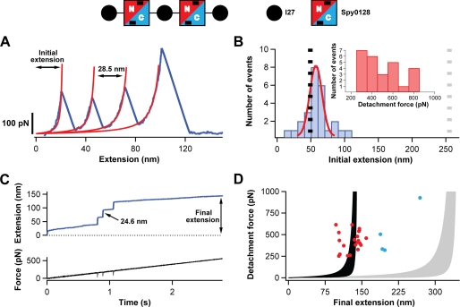
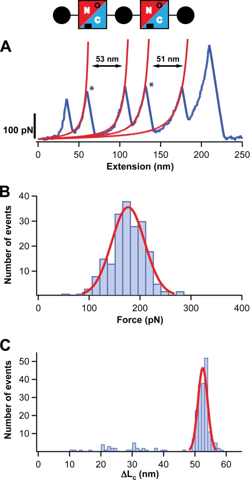
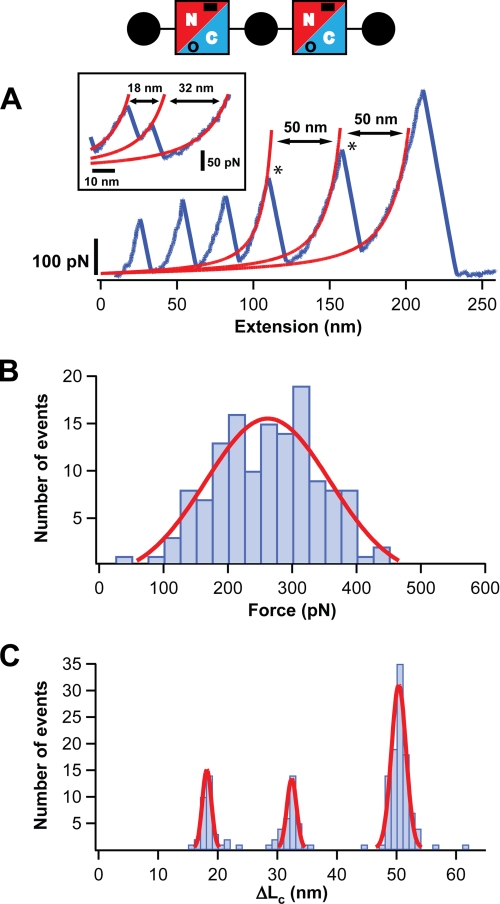
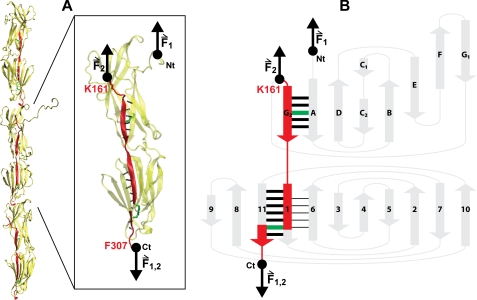
In the early stages of an infection, pathogenic bacteria use long fibrous structures known as pili as adhesive anchors for attachment to the host cells. These structures also play key roles in colony and biofilm formation. In all those processes, pili must withstand large mechanical forces. The pili of the nasty gram-positive human pathogen Streptococcus pyogenes are assembled as single, micrometer long tandem modular proteins of covalently linked repeats of pilin proteins. Here we use single molecule force spectroscopy techniques to study the mechanical properties of the major pilin Spy0128. In our studies, we engineer polyproteins containing repeats of Spy0128 flanked by the well characterized I27 protein which provides an unambiguous mechanical fingerprint. We find that Spy0128 is an inextensible protein, even when pulled at forces of up to 800 pN. We also found that this remarkable mechanical resilience, unique among the modular proteins studied to date, results from the strategically located intramolecular isopeptide bonds recently identified in the x-ray structure of Spy0128. Removal of the isopeptide bonds by mutagenesis readily allowed Spy0128 domains to unfold and extend, albeit at relatively high forces of 172 pN (N-terminal domain) or 250 pN (C-terminal domain). Our results show that in contrast to the elastic roles played by large tandem modular proteins such as titin and fibronectin, the giant pili of S. pyogenes evolved to abrogate mechanical extensibility, a property that may be crucial in the pathogenesis of this most virulent bacterium and, therefore, become the target of new therapeutic approaches against its infections.


Figures

FIGURE 1.
FIGURE 1.
Study of the extensibility of Spy0128 domains. A schematic of the protein construct used to probe the mechanical response of Spy0128 and (I27-Spy0128)2-I27 is shown. Two Spy0128 molecules (squares) were inserted between I27 domains (black circles). Spy0128 is composed of two domains (N terminus, red; C terminus, blue). Intramolecular isopeptide bonds are represented by black bars. A, typical force-extension trace (blue) showing three peaks corresponding to the unfolding of the three I27 domains in the protein construct. In red, fits to the worm-like chain model are shown. Even though the Spy0128 domains must have been subject to force, no unfolding peaks corresponding to Spy0128 are identified. B, histogram of the initial extension for traces with three I27 domains unfolding events (n = 26). The expected initial extensions if the two Spy0128 behaved as random coils (gray dotted line) or as mechanically stable domains (black dotted line) are shown. A Gaussian fit to the histogram is shown in red. Inset, histogram showing the detachment force in the force-extension experiments. C, top, typical force-ramp trace characterized by three steps marking the unfolding of the I27 modules. No steps corresponding to the Spy0128 domains were observed. Bottom, time course of the force experienced by the polyprotein. The unfolding events appear as negative spikes, reflecting the time response of the feedback system (33). D, scatter plot showing the final extension in the force-ramp experiments versus the detachment force (n = 25). The shaded regions represent the worm-like chain model of polymer elasticity (persistence lengths between 0.2 and 2.3 nm) if the Spy0128 domains remain folded (black) or, in contrast, unfold during the force-ramp protocol (gray) (28). The experimental points not following the general trend are colored in blue.
FIGURE 2.
FIGURE 2.
The N-terminal domain of Spy0128 E117A extends upon the application of force. A diagram of the protein construct employed, (I27-Spy0128-E117A)2-I27, is shown (see also legend to Fig. 1). The mutation E117A abolishes the formation of the isopeptide bond at the N-terminal domain of Spy0128 (in the diagram, this is denoted by an open circle). A, typical force-extension trace for (I27-Spy0128-E117A)2-I27 is shown in blue. The peaks marking the unfolding of the Spy0128 N-terminal domains in the polyprotein are labeled with an asterisk. The increment in contour length after the Spy0128 unfolding events is calculated from the worm-like chain fits shown in red. B, histogram of the unfolding forces for the N-terminal domain in Spy0128 E117A (n = 197). The solid line corresponds to a Gaussian fit of the experimental data. C, histogram of the ΔLc associated with the unfolding of the N-terminal domain in Spy0128 E117A (n = 214). A Gaussian fit for the data above 45 nm is shown (solid line).
FIGURE 3.
FIGURE 3.
The C-terminal domain of Spy0128 E258A extends upon the application of force. A diagram of the protein construct employed, (I27-Spy0128-E258A)2-I27, is shown (see also legend to Fig. 1). The mutation E258A abolishes the formation of the isopeptide bond at the C-terminal domain of Spy0128 (in the diagram, this is denoted by an open circle). A, typical force-extension trace for (I27-Spy0128-E258A)2-I27 is shown in blue. The peaks marking the unfolding of the Spy0128 C-terminal domains in the polyprotein are labeled with an asterisk. The increment in contour length after the Spy0128 unfolding events is calculated from the worm-like chain fits shown in red. Inset: the unfolding of the C-terminal domain through an intermediate is shown. B, histogram of the unfolding forces for the C-terminal domain in Spy0128 E258A (n = 135). The solid line corresponds to a Gaussian fit of the experimental data. C, histogram of the ΔLc associated with the unfolding of the C-terminal domain in Spy0128 E258A (n = 169). Solid lines are Gaussian fits to the data.
FIGURE 4.
FIGURE 4.
Mechanical architecture of Spy0128. A, model of the pili in S. pyogenes showing the spatial arrangement of three Spy0128 units (21). The structure of one Spy0128 monomer (PDB code: 3B2M) is enlarged. The hydrogen bonds involved in the mechanical stabilization of the domains are shown as dotted black lines. The intramolecular isopeptide bonds (Lys-36—Asn-168; Lys-179—Asn-303) are depicted in green. Pilin monomers homopolymerize to produce the pilus shaft through intermolecular isopeptide bonds between Lys-161 and the carboxyl group of Thr-311 (21). As a consequence of both the inter- and intramolecular isopeptide bonds, an axial force applied to the pilus is transmitted along the chain colored in red (instead of Thr-311, the last residue modeled in the crystal structure of Spy0128, Phe-307, is shown). The pulling directions in the polyproteins used in this work (F1) and in native pili (F2) are indicated by black arrows. VMD was used to prepare the figure (49). B, topology diagram of Spy0128. The main mechanical transitions states are represented by thick black bars. The mechanical transition state giving rise to the intermediate captured in the mechanical unfolding of Spy0128 E258A is represented by black lines between strands 1 and 6. Force pathways and intramolecular isopeptide bonds are colored as in A. The N and C termini are indicated in both panels.

References

    1. Cunningham M. W. (2008) Adv. Exp. Med. Biol. 609, 29–42 - PubMed
    1. Manetti A. G., Zingaretti C., Falugi F., Capo S., Bombaci M., Bagnoli F., Gambellini G., Bensi G., Mora M., Edwards A. M., Musser J. M., Graviss E. A., Telford J. L., Grandi G., Margarit I. (2007) Mol. Microbiol. 64, 968–983 - PubMed
    1. Abbot E. L., Smith W. D., Siou G. P., Chiriboga C., Smith R. J., Wilson J. A., Hirst B. H., Kehoe M. A. (2007) Cell Microbiol. 9, 1822–1833 - PubMed
    1. Proft T., Baker E. N. (2009) Cell Mol. Life Sci. 66, 613–635 - PMC - PubMed
    1. Craig L., Pique M. E., Tainer J. A. (2004) Nat. Rev. Microbiol. 2, 363–378 - PubMed

Publication types

LinkOut - more resources