. 2010 Apr 2;285(14):10424-33.
doi: 10.1074/jbc.M109.096966. Epub 2010 Feb 5.

Structure-function study of the N-terminal domain of exocyst subunit Sec3

Affiliations

Structure-function study of the N-terminal domain of exocyst subunit Sec3

Kyuwon Baek et al. J Biol Chem. .

Abstract

The exocyst is an evolutionarily conserved octameric complex involved in polarized exocytosis from yeast to humans. The Sec3 subunit of the exocyst acts as a spatial landmark for exocytosis through its ability to bind phospholipids and small GTPases. The structure of the N-terminal domain of Sec3 (Sec3N) was determined ab initio and defines a new subclass of pleckstrin homology (PH) domains along with a new family of proteins carrying this domain. Respectively, N- and C-terminal to the PH domain Sec3N presents an additional alpha-helix and two beta-strands that mediate dimerization through domain swapping. The structure identifies residues responsible for phospholipid binding, which when mutated in cells impair the localization of exocyst components at the plasma membrane and lead to defects in exocytosis. Through its ability to bind the small GTPase Cdc42 and phospholipids, the PH domain of Sec3 functions as a coincidence detector at the plasma membrane.


Figures

FIGURE 1.
FIGURE 1.
Sec3N folds into a PH domain. A, two perpendicular views of a superimposition of the structure of the Sec3N monomer with that of the PH domain of phospholipase Cδ (28) (PDB code: 1MAI). Phospholipase Cδ (PLCD1) is colored blue and Sec3N is colored green (helices), yellow (β-strands in front), red (β-strands in back), and cyan (variable loops 1–3). Compared with PLCD1, Sec3N contains an additional α-helix at the N terminus (α0) and two β-strands at the C terminus (magenta). The two structures superimpose with an r.m.s.d. of 2.15 Å for 87 equivalent Cα positions. B, sequence alignment of yeast Sec3N with the equivalent region of human Sec3, and the Sec3N-related proteins amisyn and maize roothairless (or rth1). PLCD1 was also aligned based on a structure superimposition. The percentage identity and similarity are indicated in the bottom right corner. Secondary structure assignment is shown above the alignment and colored according to A. Residues predicted to be important for phosphoinositide binding are highlighted (red contour). Uniprot accession codes: SEC3_YEAST (P33332), SEC3_HUMAN (Q9NV70), AMISYN_HUMAN (Q8NFX7), rth1_MAIZE (Q5YLM3), and PLCD1_RAT (P10688).
FIGURE 2.
FIGURE 2.
Sec3N forms an antiparallel dimer of PH domains. The figure shows a ribbon diagram and surface representations of the Sec3 dimer. One of the molecules of the dimer is labeled and colored according to Fig. 1, while the other is colored blue. The phosphate ions that bind in the predicted phosphoinositide-binding pockets at the distal ends of the dimer are also shown. Note that dimerization results from domain swapping of strands β8 and β9. An enlarged view shows some of the hydrophobic amino acids involved in interactions at the dimer interface.
FIGURE 3.
FIGURE 3.
Phosphoinositide-binding pocket. A, electrostatic surface representation of Sec3N near the predicted phosphoinositide-binding pocket surrounded by VL1–3. Note that the pocket is positively charged (blue color) due to the presence of various basic amino acids. B, electron density map (2FoFc, contoured at 1.5σ) in the area corresponding to the yellow square in A. The C terminus of a symmetry-related molecule binds in the phosphoinositide-binding pocket (green backbone and red electron density map). A phosphate ion, the C terminus, and the carboxylic group of Glu-240 from the symmetry-related molecule are likely to mimic protein-phosphoinositide interactions. C, using these three negative charges as reference, inositol 1,4,5-trisphosphate can be modeled in the pocket. Although it is unlikely that phosphoinositides bind precisely in this manner, this model identifies some of the amino acids most likely to participate in protein-phosphoinositide interactions.
FIGURE 4.
FIGURE 4.
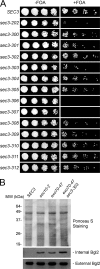
The exo70-47 sec3-303 double mutant shows growth and secretion defects. A, various sec3 mutant alleles were introduced into the exo70-47 mutant strain, in which the chromosomal copy of EXO70 was deleted and replaced by the HIS3 gene. The exo70-47 mutant was expressed under the EXO70 promoter in a CEN TRP1 plasmid, and the cells were supplemented with an EXO70 balancer on a CEN URA3 plasmid. The cells were grown at 25 °C on 5-FOA-containing medium, which triggers the elimination of the EXO70 balancer plasmids and subsequent generation of the exo70-47 sec3 double mutants (right panel). Controls were grown on medium without 5-FOA (left panel). sec3-303 and several other mutants showed different degrees of synthetic growth defects with exo70-47. sec3-306 and sec3-307 were synthetically lethal with exo70-47. B, EXO70, exo70-47, and exo70-47 sec3-303 cells were grown to early log phase. The sec10-2 mutant cells were grown to early log phase and then shifted to 37 °C for 1 h. The internal and external pools of Bgl2 were analyzed by Western blot using an anti-Bgl2 antibody. The exo70-47 and sec10-2 mutants showed defects in Bgl2 secretion. The sec3-303 exo70-47 double mutant was more defective in Bgl2 secretion than the exo70-47 single mutant. Ponceau S staining is shown to indicate equal protein loading.
FIGURE 5.
FIGURE 5.
Localization of exocyst components in exo70-47 and exo70-47 sec3-303 mutant cells. A, the exocyst components in the exo70-47 single mutant and the exo70-47 sec3-303 double mutant cells were GFP-tagged by chromosomal integration. GFP-tagged Sec3, Sec5, Sec8, and Exo84 were localized to the bud tip in the single mutants. In the double mutants, sec3-303-GFP was diffused throughout the cell, while the rest of the GFP-tagged exocyst proteins were localized to the entire bud instead of the bud tip. GFP-Sec6 was diffused in the entire bud in both single and double mutants. B and C, representative histograms depicting the fluorescence intensity along the bud and into the cell body (along the white line) in single (exo70-47) and double (exo70-47 sec3-303) mutants expressing Sec3-GFP or sec3-303-GFP (B) or Exo84-GFP (C) (see insets).

Similar articles

Cited by

  • Beyond symmetry-breaking: competition and negative feedback in GTPase regulation.
    Wu CF, Lew DJ. Wu CF, et al. Trends Cell Biol. 2013 Oct;23(10):476-83. doi: 10.1016/j.tcb.2013.05.003. Epub 2013 May 31. Trends Cell Biol. 2013. PMID: 23731999 Free PMC article. Review.
  • Integrative structure and function of the yeast exocyst complex.
    Ganesan SJ, Feyder MJ, Chemmama IE, Fang F, Rout MP, Chait BT, Shi Y, Munson M, Sali A. Ganesan SJ, et al. Protein Sci. 2020 Jun;29(6):1486-1501. doi: 10.1002/pro.3863. Epub 2020 May 1. Protein Sci. 2020. PMID: 32239688 Free PMC article.
  • Cell Polarity in Yeast.
    Chiou JG, Balasubramanian MK, Lew DJ. Chiou JG, et al. Annu Rev Cell Dev Biol. 2017 Oct 6;33:77-101. doi: 10.1146/annurev-cellbio-100616-060856. Epub 2017 Aug 7. Annu Rev Cell Dev Biol. 2017. PMID: 28783960 Free PMC article. Review.
  • The exocyst in context.
    Meek S, Hernandez AC, Oliva B, Gallego O. Meek S, et al. Biochem Soc Trans. 2024 Oct 30;52(5):2113-2122. doi: 10.1042/BST20231401. Biochem Soc Trans. 2024. PMID: 39377315 Free PMC article. Review.
  • Exorcising the exocyst complex.
    Heider MR, Munson M. Heider MR, et al. Traffic. 2012 Jul;13(7):898-907. doi: 10.1111/j.1600-0854.2012.01353.x. Epub 2012 Apr 8. Traffic. 2012. PMID: 22420621 Free PMC article. Review.

References

    1. Novick P., Field C., Schekman R. (1980) Cell 21, 205–215 - PubMed
    1. TerBush D. R., Novick P. (1995) J. Cell Biol. 130, 299–312 - PMC - PubMed
    1. Hsu S. C., Ting A. E., Hazuka C. D., Davanger S., Kenny J. W., Kee Y., Scheller R. H. (1996) Neuron 17, 1209–1219 - PubMed
    1. Munson M., Novick P. (2006) Nat. Struct. Mol. Biol. 13, 577–581 - PubMed
    1. He B., Guo W. (2009) Curr. Opin. Cell Biol. 21, 537–542 - PMC - PubMed

Publication types

MeSH terms

Substances