. 2010 Apr 9;285(15):11188-96.
doi: 10.1074/jbc.M109.097675. Epub 2010 Feb 5.

Molecular interaction and functional regulation of ClC-3 by Ca2+/calmodulin-dependent protein kinase II (CaMKII) in human malignant glioma

Affiliations

Molecular interaction and functional regulation of ClC-3 by Ca2+/calmodulin-dependent protein kinase II (CaMKII) in human malignant glioma

Vishnu Anand Cuddapah et al. J Biol Chem. .

Abstract

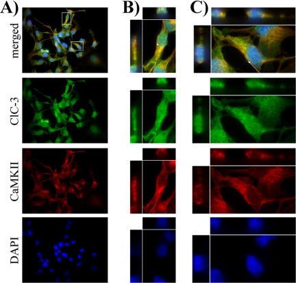
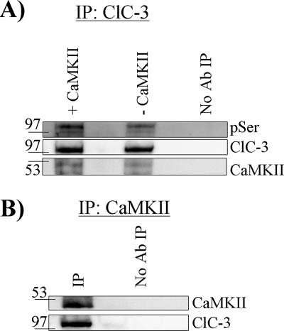
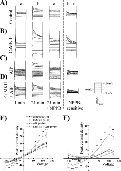
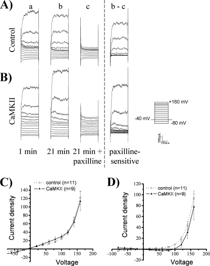
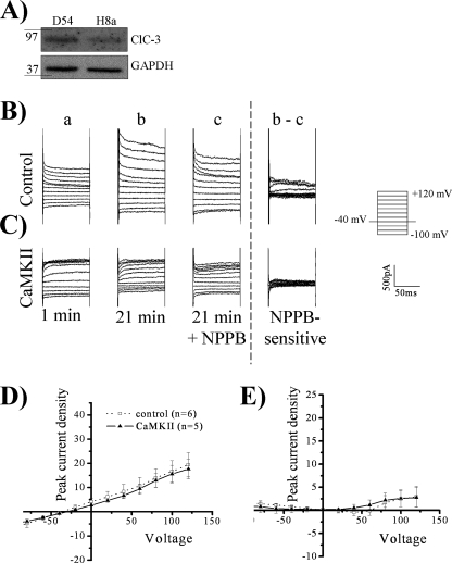
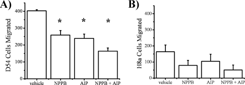
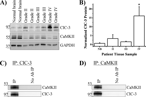
Glioblastoma multiforme is the most common and lethal primary brain cancer in adults. Tumor cells diffusely infiltrate the brain making focal surgical and radiation treatment challenging. The invasion of glioma cells into normal brain is facilitated by the activity of ion channels aiding dynamic regulation of cell volume. Recent studies have specifically implicated ClC-3, a voltage-gated chloride channel, in this process. However, the interaction between ClC-3 activity and cell movement is poorly understood. Here, we demonstrate that ClC-3 is highly expressed on the plasma membrane of human glioma cells where its activity is regulated through phosphorylation via Ca(2+)/calmodulin-dependent protein kinase II (CaMKII). Intracellular infusion of autoactivated CaMKII via patch pipette enhanced chloride currents 3-fold, and this regulation was inhibited by autocamtide-2 related inhibitory peptide, a CaMKII-specific inhibitor. CaMKII modulation of chloride currents was also lost upon stable small hairpin RNA knockdown of ClC-3 channels indicating a specific interaction of ClC-3 and CaMKII. In ClC-3-expressing cells, inhibition of CaMKII reduced glioma invasion to the same extent as direct inhibition of ClC-3. The importance of the molecular interaction of ClC-3 and CaMKII is further supported by our finding that CaMKII co-localizes and co-immunoprecipitates with ClC-3. ClC-3 and CaMKII also co-immunoprecipitate in tissue biopsies from patients diagnosed with grade IV glioblastoma. These tumor samples show 10-fold higher ClC-3 protein expression than nonmalignant brain. These data suggest that CaMKII is a molecular link translating intracellular calcium changes, which are intrinsically associated with glioma migration, to changes in ClC-3 conductance required for cell movement.


Figures

FIGURE 1.
FIGURE 1.
Human glioma cells express ClC-3 on the plasma membrane. The 1st row contains representative merged images, with examples of ClC-3 and cortical actin co-localization indicating ClC-3 expression on the plasma membrane. The 2nd row demonstrates ClC-3 immunolabeling (green) throughout the cell. The 3rd row shows phalloidin binding of actin (red), including cortical actin on the plasma membrane. The 4th row is the DAPI nuclear stain (blue). A, ×60 view of a field of glioma cells. Boxes demarcate zoomed cells seen in B and C. B and C, digital zooms of individual cells. Arrows point to cross-sections of cells seen in three-view through the entire 10-μm imaging plane.
FIGURE 2.
FIGURE 2.
ClC-3 and CaMKII co-localize in human glioma cells. The 1st row contains representative merged images, with examples of ClC-3 and CaMKII co-localization. The 2nd row demonstrates ClC-3 immunolabeling (green) throughout the cell. The 3rd row shows CaMKII immunolabeling (red). The 4th row is the DAPI nuclear stain (blue). A, ×60 view of a field of glioma cells. Boxes demarcate zoomed cells seen in B and C. B and C, digital zooms of individual cells. Arrows point to cross-sections of cells seen in three-view through the entire 10-μm imaging plane.
FIGURE 3.
FIGURE 3.
CaMKII phosphorylates and co-immunoprecipitates with ClC-3. A, after immunoprecipitation (IP) of ClC-3, CaMKII phosphorylated ClC-3 as seen by enhanced binding of phosphoserine (pSer) antibody (Ab). Equal amounts of ClC-3 were pulled down, and CaMKII was detected. B, after immunoprecipitation of CaMKII, ClC-3 was detected only when the precipitating antibody was included. Blots are representative of n ≥3 experiments.
FIGURE 4.
FIGURE 4.
ClC-3 current is activated by CaMKII. Using whole-cell patch clamp electrophysiology, human glioma cells were held at −40 mV and stepped from −100 mV to +120 mV. For the representative traces, the 1st column (a) is basal current at 1 min, and the 2nd column (b) is current at 21 min. At 21 min, NPPB was added as seen in the 3rd column (c), and the NPPB-sensitive current (bc) is depicted in the 4th column. A, control condition. B, activated CaMKII included in the pipette solution leading to an enhancement of chloride current. C, AIP, a CaMKII inhibitor, is included in the pipette solution. D, activated CaMKII and AIP are included in the pipette solution leading to a loss of CaMKII-mediated current enhancement. E, peak whole-cell current density corresponding to b. F, peak NPPB-sensitive current density corresponding to bc. p values indicate significance of CaMKII versus CaMKII + AIP conditions (*, p < 0.05; **, p < 0.01; ***, p < 0.001; Tukey Kramer). n = 10–11.
FIGURE 5.
FIGURE 5.
BK channels are not activated by CaMKII. Using whole-cell patch clamp electrophysiology, human glioma cells were held at −40 mV and stepped from −80 mV to +160 mV. For the representative traces, the 1st column (a) is basal current at 1 min, and the 2nd column (b) is current at 21 min. At 21 min, paxilline was added as seen in the 3rd column (c), and the paxilline-sensitive current (bc) is depicted in the 4th column. A, control condition. B, activated-CaMKII included in the pipette solution does not lead to a change in current. C, whole-cell current density corresponding to b. D, paxilline-sensitive current density corresponding to bc is not significantly different from control. n = 9–11.
FIGURE 6.
FIGURE 6.
CaMKII does not activate a chloride current after ClC-3 knockdown. A, ClC-3 expression is decreased in H8a cells expressing ClC-3 small hairpin RNA relative to glyceraldehyde-3-phosphate dehydrogenase (GAPDH) loading control, and CaMKII expression is not altered. B–E, using whole-cell patch clamp electrophysiology, human glioma cells were held at −40 mV and stepped from −100 mV to +120 mV. For the representative traces, the 1st column (a) is basal current at 1 min, and the 2nd column (b) is current at 21 min. At 21 min, NPPB was added as seen in the 3rd column (c), and the NPPB-sensitive current (bc) is depicted in the 4th column. B, control condition. C, activated CaMKII included in the pipette solution does not lead to an enhancement of chloride current. D, peak whole-cell current density corresponding to b. E, peak NPPB-sensitive current density corresponding to b and c. n = 5–6.
FIGURE 7.
FIGURE 7.
ClC-3 or CaMKII inhibition leads to an attenuation in Transwell migration. A, representative images of Transwell migration of D54 and H8a (ClC-3 knock-down) cells after drug treatment. B, NPPB, AIP, and NPPB + AIP decrease D54 cell migration. C, ClC-3 knockdown leads to a decrease in migration that is not altered after drug treatment. (*, p < 0.05; Tukey Kramer). n = 3.
FIGURE 8.
FIGURE 8.
ClC-3 is overexpressed in grade IV human glioma tissue and associated with CaMKII. A, representative blot demonstrating ClC-3 and CaMKII immunoreactivity in normal brain, grade II, grade III, and grade IV human biopsy samples. B, quantification of ClC-3 expression normalized to normal brain reveals overexpression in grade IV samples. (*, p < 0.05; Tukey Kramer). (Normal brain, n = 3; grade II, n = 4; grade III, n = 3; and grade IV, n = 4.) C, after immunoprecipitation of ClC-3 from a grade IV glioblastoma patient biopsy, CaMKII was detected only when the precipitating antibody was included. D, after immunoprecipitation of CaMKII from a grade IV glioblastoma patient biopsy, ClC-3 was detected only when the precipitating antibody was included. Blots are representative of n ≥ 3 experiments.

Similar articles

Cited by

  • ANO1 as a marker of oral squamous cell carcinoma and silencing ANO1 suppresses migration of human SCC-25 cells.
    Li Y, Zhang J, Hong S. Li Y, et al. Med Oral Patol Oral Cir Bucal. 2014 Jul 1;19(4):e313-9. doi: 10.4317/medoral.19076. Med Oral Patol Oral Cir Bucal. 2014. PMID: 24316695 Free PMC article.
  • Combating malignant astrocytes: Strategies mitigating tumor invasion.
    Umans RA, Sontheimer H. Umans RA, et al. Neurosci Res. 2018 Jan;126:22-30. doi: 10.1016/j.neures.2017.09.010. Epub 2017 Oct 18. Neurosci Res. 2018. PMID: 29054465 Free PMC article. Review.
  • Integrated Proteogenomic Characterization across Major Histological Types of Pediatric Brain Cancer.
    Petralia F, Tignor N, Reva B, Koptyra M, Chowdhury S, Rykunov D, Krek A, Ma W, Zhu Y, Ji J, Calinawan A, Whiteaker JR, Colaprico A, Stathias V, Omelchenko T, Song X, Raman P, Guo Y, Brown MA, Ivey RG, Szpyt J, Guha Thakurta S, Gritsenko MA, Weitz KK, Lopez G, Kalayci S, Gümüş ZH, Yoo S, da Veiga Leprevost F, Chang HY, Krug K, Katsnelson L, Wang Y, Kennedy JJ, Voytovich UJ, Zhao L, Gaonkar KS, Ennis BM, Zhang B, Baubet V, Tauhid L, Lilly JV, Mason JL, Farrow B, Young N, Leary S, Moon J, Petyuk VA, Nazarian J, Adappa ND, Palmer JN, Lober RM, Rivero-Hinojosa S, Wang LB, Wang JM, Broberg M, Chu RK, Moore RJ, Monroe ME, Zhao R, Smith RD, Zhu J, Robles AI, Mesri M, Boja E, Hiltke T, Rodriguez H, Zhang B, Schadt EE, Mani DR, Ding L, Iavarone A, Wiznerowicz M, Schürer S, Chen XS, Heath AP, Rokita JL, Nesvizhskii AI, Fenyö D, Rodland KD, Liu T, Gygi SP, Paulovich AG, Resnick AC, Storm PB, Rood BR, Wang P; Children’s Brain Tumor Network; Clinical Proteomic Tumor Analysis Consortium. Petralia F, et al. Cell. 2020 Dec 23;183(7):1962-1985.e31. doi: 10.1016/j.cell.2020.10.044. Epub 2020 Nov 25. Cell. 2020. PMID: 33242424 Free PMC article.
  • Ion channels in glioblastoma.
    Molenaar RJ. Molenaar RJ. ISRN Neurol. 2011;2011:590249. doi: 10.5402/2011/590249. Epub 2011 Nov 29. ISRN Neurol. 2011. PMID: 22389824 Free PMC article.
  • The emerging role of CaMKII in cancer.
    Wang YY, Zhao R, Zhe H. Wang YY, et al. Oncotarget. 2015 May 20;6(14):11725-34. doi: 10.18632/oncotarget.3955. Oncotarget. 2015. PMID: 25961153 Free PMC article. Review.

References

    1. Wen P. Y., Kesari S. (2008) N. Engl. J. Med. 359, 492–507 - PubMed
    1. Farin A., Suzuki S. O., Weiker M., Goldman J. E., Bruce J. N., Canoll P. (2006) Glia 53, 799–808 - PubMed
    1. Bernstein J. J., Goldberg W. J., Laws E. R., Jr. (1989) J. Neurosci. Res. 22, 134–143 - PubMed
    1. Sontheimer H. (2008) Exp. Biol. Med. 233, 779–791 - PMC - PubMed
    1. Liu X., Chang Y., Reinhart P. H., Sontheimer H., Chang Y. (2002) J. Neurosci. 22, 1840–1849 - PMC - PubMed

Publication types

MeSH terms