. 2010 Apr 16;285(16):12344-54.
doi: 10.1074/jbc.M110.106856. Epub 2010 Feb 5.

The Rho guanine nucleotide exchange factor AKAP13 (BRX) is essential for cardiac development in mice

Affiliations

The Rho guanine nucleotide exchange factor AKAP13 (BRX) is essential for cardiac development in mice

Chantal M Mayers et al. J Biol Chem. .

Abstract

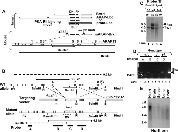
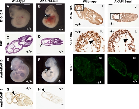
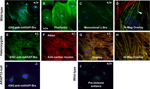
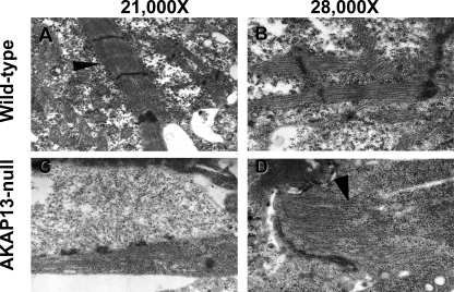
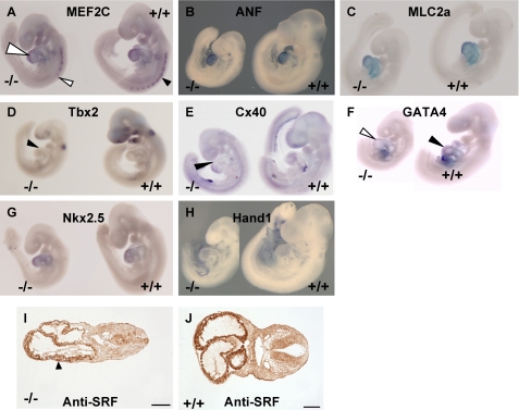
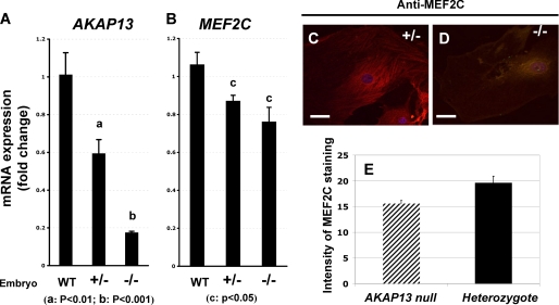
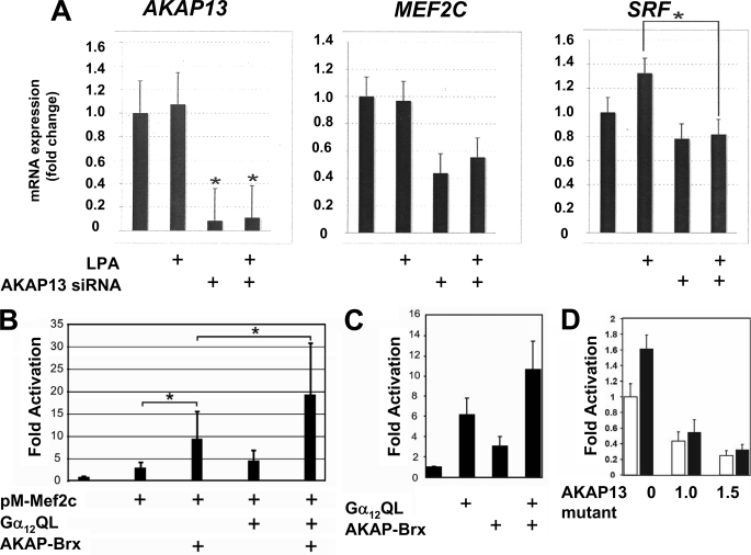
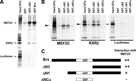
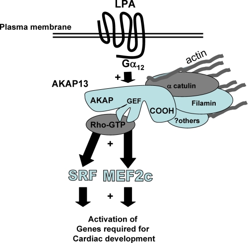
A fundamental biologic principle is that diverse biologic signals are channeled through shared signaling cascades to regulate development. Large scaffold proteins that bind multiple proteins are capable of coordinating shared signaling pathways to provide specificity to activation of key developmental genes. Although much is known about transcription factors and target genes that regulate cardiomyocyte differentiation, less is known about scaffold proteins that couple signals at the cell surface to differentiation factors in developing heart cells. Here we show that AKAP13 (also known as Brx-1, AKAP-Lbc, and proto-Lbc), a unique protein kinase A-anchoring protein (AKAP) guanine nucleotide exchange region belonging to the Dbl family of oncogenes, is essential for cardiac development. Cardiomyocytes of Akap13-null mice had deficient sarcomere formation, and developing hearts were thin-walled and mice died at embryonic day 10.5-11.0. Disruption of Akap13 was accompanied by reduced expression of Mef2C. Consistent with a role of AKAP13 upstream of MEF2C, Akap13 siRNA led to a reduction in Mef2C mRNA, and overexpression of AKAP13 augmented MEF2C-dependent reporter activity. The results suggest that AKAP13 coordinates Galpha(12) and Rho signaling to an essential transcription program in developing cardiomyocytes.


Figures

FIGURE 1.
FIGURE 1.
Diagram of murine Akap13 gene and deletion strategy. A, the human AKAP13 gene encodes a 5.3-kb brx-1 transcript and an 8.5- and 10-kb transcript. Shown is the murine AKAP-Brx transcript and genomic intron-exon structure with DH and pleckstrin homology (PH) regions as indicated. Binding sites of antiserum 4362 and anti-BRX monoclonal antibody mab are shown. Exons are numbered consecutively beginning at the GEF region. B, partial restriction map of the wild-type murine Akap13 GEF region, the targeting vector, and the mutant Akap13 allele. Shown are the neomycin cassette (striped) insertion with deletion of part of exon 4, the entire exon 5, and 5398 bp of flanking genomic sequence. Dark gray, exons; white, introns. Probes A–D are shown as black bars. Double-headed solid arrows, restriction fragments generated by BamHI digestion of wild-type (3.9 kb) and mutant alleles (4.8 kb) with 5′-probe B (black bar). The BamHI site deleted in the mutant allele is boxed. Double-headed dashed arrows depict restriction fragments for EcoRV digestion probed with 5′-probe A. The inserted EcoRV site is shown as a dashed box. C, Southern analysis of targeted BamHI-digested embryonic stem cell DNA (lanes 1 and 2) or tail DNA (lanes 3 and 4) with 5′-probe B revealed a recombinant 4.8 kb band and 3.9 kb wild-type bands (open arrowhead) in heterozygous (Ht) embryonic stem cells and mice. D, RT-PCR amplification of mRNA from E9.0–9.5 embryos. 90–99 ng of cDNA product from embryos was amplified with 33 pmol of primers for either murine Akap13 (odd lanes) or glyceraldehyde-3-phosphate dehydrogenase (even lanes). Murine cDNA was used as a positive control (lane 9). The expected 550 bp band was present in lanes 1, 3, and 7, consistent with the heterozygous genotype (black triangle). An Akap13 transcript was not detectable in mRNA harvested from an embryo homozygous for the Akap13 null allele (lane 5). Results were confirmed in four independent experiments. E, Northern analysis of a multi-tissue mRNA blot using an EcoRI-EcoRI fragment from the 3′-region of the murine Akap13 cDNA. A 9.5–10.0 kb band was detected in RNA harvested from heart tissues.
FIGURE 2.
FIGURE 2.
Phenotype of Akap13-null mutant embryos. Sibling embryos are shown in A and B, C and D, E and F, and I and J. A, dissecting microscope view of wild-type embryo at E10–10.5 (bar for comparison). B, mutant sibling embryo with an enlarged heart and pericardial effusion (white arrow). Somite development of mutant embryos was within two somite pairs of wild-type embryos at E9.0–9.5. C, transverse section (hematoxylin and eosin (H&E)) through wild-type embryo at E9.5–10.0. V, ventricle. Bar, 100 μm. Magnification was ×10. D, transverse section through mutant sibling embryo. V, ventricle. Note the thin myocardium. E, whole mount immunohistochemical staining of wild-type embryo using 4362 antiserum. AKAP13 protein was present in heart, limb bud, first brachial arch, and forebrain. F, whole mount immunohistochemical staining of mutant embryo. No staining was observed. The developing heart shows a pericardial effusion and a constriction corresponding to the primary ring (arrowhead). G, transverse section of heterozygous embryo E10.0–10.5 reacted with 1:1000 antiserum 4362. Strong brown staining was present in the developing heart identical to wild-type embryos. Bar, 50 μm. H, transverse section of mutant embryo stained with 1:1000 dilution of antiserum 4362. Intensity of staining of Akap13-null embryos resembled the intensity of staining observed in wild-type embryos reacted with preimmune serum (not shown). Shown is transverse section of wild-type embryo (I) or mutant sibling embryo (J) stained with antiserum against Ki67. K and L, ×40 magnification of the section boxed in I and J, respectively. A reduction in cardiomyocyte number in the developing ventricle was noted at ×40 magnification (compare black arrowheads in K and L). M and N, TUNEL assay of cardiac tissues at E9.5 from either a wild-type (M), or Akap13-null (N) embryos.
FIGURE 3.
FIGURE 3.
Localization of AKAP13 to the cytoskeleton. A, confocal image of primary culture of cardiomyocytes from a wild-type (+/+) embryos at E9.0–9.5 plated on collagen and stained with antiserum 4362 directed against AKAP13 and DAPI. B, confocal image of cardiomyocytes of wild-type embryo plated on collagen and stained with FITC-phalloidin and DAPI. C, wild-type cardiomyocytes stained with a monoclonal antibody directed against AKAP13 and DAPI (×40, oil). D, high magnification; overlay (+/+) cardiomyocytes stained with 4362 antiserum (green) and actin (red). The yellow signal (arrowhead) shows co-localization of AKAP13 and actin. E, heterozygote cardiomyocytes plated on fibronectin stained with antiserum 4362 and DAPI. Intensity of staining from cardiomyocytes from heterozygous mice resembled that in cells from wild-type mice. Cells plated on fibronectin showed more robust actin-myosin filaments. F, heterozygote cardiomyocytes plated on fibronectin stained with monoclonal antibody specific for heavy chain cardiac myosin (ab50967) (×40, oil). G, overlay of heterozygote cell in E and F. The yellow signal shows co-localization of AKAP13 and cardiac myosin (×40, oil). H, high magnification overlay showing myosin bands. I, cardiomyocytes from an Akap13 null embryo at E9.0–9.5 stained with 4362 antiserum showed only background levels of signal. J, wild-type cardiomyocytes stained with preimmune serum showed no staining of cytoskeleton (×40, oil).
FIGURE 4.
FIGURE 4.
Altered sarcomere development in Akap13-null mice. A, transmission electron micrograph from developing heart at E9.0–9.5 of wild-type mice exhibited normal sarcomere structure. Arrowhead, Z-disc. B, ×28,000 magnification of sarcomere from wild-type mouse. C, transmission electron micrograph of mutant heart at E9.0–9.5 showed few sarcomere structures and structures lacking distinct Z-discs. D, ×28,000 magnification of mutant sarcomere. Note that the sarcomere ended blindly (black arrowhead).
FIGURE 5.
FIGURE 5.
Altered expression of heart markers in Akap13-null embryos. A–H, whole mount in situ hybridization of sibling embryos E9–9.5 with either a mutant genotype (−/−) or a wild-type (+/+) genotype. Each panel is a representative of three independent experiments with sibling mutant embryos. A, staining for Mef2C mRNA was reduced in AKAP13-null embryos (left) compared with wild-type littermates. Reduced staining was noted in somites (arrowheads) and heart. B, Anf transcripts did not differ between wild-type littermates and mutant embryos. C, Mlc2a transcripts were similarly expressed. D, Tbx2 staining was reduced in Akap13-null embryos (black triangle) compared with wild-type littermates. E, connexin 40 (Cx40) transcripts were localized to the extreme anterior ventricle in the Akap13-null embryos (black triangle) compared with wild-type littermates. F, Gata-4 transcripts were slightly reduced, in developing hearts of Akap13−/− embryos (open triangle) compared with wild-type embryos (black triangle). G, staining for Nkx2.5 transcripts was similar in −/− embryos compared with wild-type littermates. H, Hand1 transcripts in Akap13−/− embryos were comparable with wild-type littermates. I, transverse section of mutant embryo reacted with 1:500 antiserum directed against SRF. SRF protein was reduced in the developing hearts of mutant embryos. Bar, 100 μm. J, transverse section of wild-type embryo reacted with 1:500 antiserum directed against SRF. Strong staining is observed in myocardium. Bar, 100 μm.
FIGURE 6.
FIGURE 6.
AKAP13 is required for expression of normal levels of MEF2C. A, quantification of Akap13 mRNA levels in developing hearts at E9.0 using real-time RT-PCR. Levels of Akap13 transcripts in the hearts were compared in embryos that were wild type (WT), haploinsufficient, or homozygous null for the Akap13 gene. Transcript levels in the −/− embryos resembled background. y axis, relative expression. B, quantification of Mef2C transcript levels in embryonic hearts at E9.0. Genotypes on the x axis refer to Akap13 genotypes. Mef2C transcripts in hearts of −/− embryos were reduced 25–30% from the levels present in wild-type mice. C, representative cardiomyocyte from heterozygote embryo plated on collagen and stained for MEF2C (E-17) and Alexa-594. D, representative cardiomyocyte from Akap13-null mouse stained for MEF2C and Alexa-594. E, quantification of confocal staining intensity for MEF2C from heterozygote or Akap13-null cardiomyocytes. y axis, pixels/area; error bars, ±S.E.; p < 0.05.
FIGURE 7.
FIGURE 7.
AKAP13 augments MEF2C-dependent gene activation via Gα12. A, endogenous AKAP13 is required for optimal MEF2C expression. H9C2 cells were transfected with control or Akap13 siRNA and treated with LPA as indicated. Total RNA was purified, and amounts of Akap13, Mef2C, or Srf were determined by real-time RT-PCR. Relative expression levels of mRNA are shown as -fold induction over base line. Results were confirmed in two separate experiments. Error bars, S.E. Mef2C mRNA levels were significantly reduced. *, p < 0.01. B, H9C2 cells were grown to 50% confluence on 12-well plates and transfected with 1.0 μg of Gal4E1b-Luc, 600 ng of pM-MEF2C, 50 ng of Gα12QL, and 1.2 μg of AKAP13 expression vectors as indicated. Following overnight incubation, cells were lysed, and luciferase assays were performed. Results are from three independent experiments. *, p < 0.05. Basal levels of reporter activity were not increased by AKAP13 alone (supplemental Fig. S5). C, transfection of serum-starved COS-7 cells (COS-7 cells were chosen because of low levels of endogenous AKAP13) with an SRE-luciferase reporter, 500 ng of an AKAP-BRX expression vector, and 400 ng of a Gα12QL expression vector, as indicated. y axis, -fold activation. Error bars, ±S.E. D, an AKAP13 mutant lacking the GEF region functions in a dominant-negative manner to inhibit activation of an SRE reporter. Shown is transient transfection of OVCAR-3 cells (shown here because of high levels of endogenous AKAP13) with 1.0 μg of SRE-luciferase reporter and the indicated amounts of an AKAP13 expression construct lacking the GEF region (AKAP13 mutant) in the presence (solid bars) or absence (open bars) of 1 μm LPA. Results of two experiments repeated in quadruplicate are shown. Error bars, ±S.E. The addition of the AKAP13 mutant lacking the GEF region inhibited SRF activation by endogenous levels of AKAP13.
FIGURE 8.
FIGURE 8.
The carboxyl region of AKAP13 (BRX) binds to MEF2C. A, 35S-labeled MEF2C and RXR bound avidly to GST fusion proteins of BRX or a GST fusion protein containing only the carboxyl region (ΔN3). B, 35S-labeled MEF2C and RXR bound avidly to BRX mutants (black arrowhead). The carboxyl-terminal region of BRX was sufficient for MEF2C binding. The 35S-labeled RXRβ or luciferase was used and positive and negative controls, respectively. C, schematic diagram of BRX mutants (see Ref. 1). GST-BRX includes amino acids 47–1428. ΔN1 contains amino acids 236–1428 of BRX. ΔN3 includes amino acids 960–1428. ΔNCα contains amino acids 527–950.
FIGURE 9.
FIGURE 9.
Model of AKAP13 action in developing heart cells. AKAP13 is activated by extracellular molecules, such as LPA, through Gα12, leading to augmentation of MEF2C and SRF activation. AKAP13 is co-localized with actin in cardiomyocytes, possibly through previously reported interactions with α-catulin (18, 28) and filamins A and B (18, 43). MEF2C and SRF are known to play critical roles in cardiomyocyte differentiation (32, 37).

Similar articles

Cited by

References

    1. Rubino D., Driggers P., Arbit D., Kemp L., Miller B., Coso O., Pagliai K., Gray K., Gutkind S., Segars J. (1998) Oncogene 16, 2513–2526 - PubMed
    1. Miller B. T., Rubino D. M., Driggers P. H., Haddad B., Cisar M., Gray K., Segars J. H. (2000) Am. J. Obstet. Gynecol. 182, 286–295 - PubMed
    1. Diviani D., Soderling J., Scott J. D. (2001) J. Biol. Chem. 276, 44247–44257 - PubMed
    1. Toksoz D., Williams D. A. (1994) Oncogene 9, 621–628 - PubMed
    1. Sterpetti P., Hack A. A., Bashar M. P., Park B., Cheng S. D., Knoll J. H., Urano T., Feig L. A., Toksoz D. (1999) Mol. Cell. Biol. 19, 1334–1345 - PMC - PubMed

Publication types

MeSH terms

Substances