. 2010 Apr;298(4):L600-6.
doi: 10.1152/ajplung.00122.2009. Epub 2010 Feb 5.

Hypoxia-induced proliferation of human pulmonary microvascular endothelial cells depends on epidermal growth factor receptor tyrosine kinase activation

Affiliations

Hypoxia-induced proliferation of human pulmonary microvascular endothelial cells depends on epidermal growth factor receptor tyrosine kinase activation

Inimary T Toby et al. Am J Physiol Lung Cell Mol Physiol. 2010 Apr.

Abstract

We hypothesized that hypoxia would activate epidermal growth factor receptor (EGFR) tyrosine kinase, leading to increased arginase expression and resulting in proliferation of human pulmonary microvascular endothelial cell (hPMVEC). To test this hypothesis, hPMVEC were incubated in normoxia (20% O(2), 5% CO(2)) or hypoxia (1% O(2), 5% CO(2)). Immunoblotting for EGFR and proliferating cell nuclear antigen was done, and protein levels of both total EGFR and proliferating cell nuclear antigen were greater in hypoxic hPMVEC than in normoxic hPMVEC. Furthermore, hypoxic hPMVEC had greater levels of EGFR activity than did normoxic hPMVEC. Hypoxic hPMVEC had a twofold greater level of proliferation compared with normoxic controls, and this increase in proliferation was prevented by the addition of AG-1478 (a pharmacological inhibitor of EGFR). Immunoblotting for arginase I and arginase II demonstrated a threefold induction in arginase II protein levels in hypoxia, with little change in arginase I protein levels. The hypoxic induction of arginase II protein was prevented by treatment with AG-1478. Proliferation assays were performed in the presence of arginase inhibitors, and hypoxia-induced proliferation was also prevented by arginase inhibition. Finally, treatment with an EGFR small interfering RNA prevented hypoxia-induced proliferation and urea production. These findings demonstrate that hypoxia activates EGFR tyrosine kinase, leading to arginase expression and thereby promoting proliferation in hPMVEC.


Figures

Fig. 1.
Fig. 1.
Time course of human pulmonary microvascular endothelial cell (hPMVEC) proliferation in normoxia and hypoxia. hPMVEC were seeded in six-well plates, with 85,000 cells in each well. After 1, 2, 3, 4, or 5 days, cells were harvested and counted for viable cell numbers using Trypan blue exclusion. The number of viable hPMVEC was greater in hypoxia (○) than in normoxia (●) on days 1, 2, 4, and 5. The growth rates of both the normoxic and hypoxic cells could be fit using linear regression. The equation for the fit of the normoxic hPMVEC was y = 27,667x + 151,667 (r = 0.94, P < 0.05), and the equation for the fit of the hypoxic hPMVEC was y = 81,333x + 253,333 (r = 0.94, P < 0.05). Both normoxic and hypoxic hPMVEC showed a positive correlation between viable cell number and day in culture. The slope for the hypoxic hPMVEC was greater (P < 0.05) than the slope for the normoxic hPMVEC by covariance analysis.
Fig. 2.
Fig. 2.
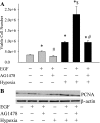
Treatment of hPMVEC with epidermal growth factor (EGF) increased cell proliferation, and this EGF-induced increase in proliferation was prevented by EGF receptor (EGFR) inhibition. A: each well in a six-well plate was seeded with 90,000 cells. The cells were treated with vehicle, EGF (50 ng/ml), or EGF + AG-1478 (30 nM) and incubated for 48 h in either normoxia (shaded bars) or hypoxia (solid bars). The number of viable cells was then counted using Trypan blue exclusion. *Different from vehicle-treated normoxia, P < 0.05. $Hypoxia EGF treated different from vehicle-treated hypoxia, P < 0.05. #EGF + AG-1478 different from EGF alone with the same condition, P < 0.05. B: proliferating cell nuclear antigen (PCNA) expression is increased in hypoxia. A representative immunoblot for PCNA expression in hPMVECs treated with either vehicle, EGF (50 ng/ml), or EGF + AG-1478 (30 nM) and then placed either in normoxia or hypoxia for 24 h is shown. The blot was stripped and reprobed with an antibody against β-actin. The experiment was repeated three times.
Fig. 3.
Fig. 3.
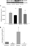
Total EGFR protein levels increase in hypoxia. A: hPMVEC were treated with either AG-1478 (30 nM) or vehicle (n = 4 in each group) and incubated in either normoxia or hypoxia for 24 h. The cell lysates were used in immunoblot analysis for total EGFR protein levels. B: EGFR mRNA levels increase in hypoxia. In a second set of exposures, RNA was collected and utilized for real-time PCR using primers for human EGFR. EGFR expression was normalized to 18S rRNA using the ΔΔCT method and expressed as fold change from normoxia. AU, arbitrary units. *Vehicle-treated hypoxia is different from vehicle-treated normoxia, P < 0.05. #AG-1478 is different from vehicle treated for the same condition, P < 0.05.
Fig. 4.
Fig. 4.
Hypoxic hPMVEC lysates have greater AG-1478-inhibitable tyrosine kinase activity than lysates from normoxic hPMVEC. Cell lysates from hPMVEC incubated in either normoxia or hypoxia for 24 h were used in a tyrosine kinase activity assay. Duplicate samples were treated with either vehicle or AG-1478, and the difference between the absorbance of the vehicle-treated well and the AG-1478-treated well was referred to as the AG-1478-inhibitable absorbance and is a measure of EGFR tyrosine kinase activity. *Hypoxic hPMVEC lysates different from normoxic hPMVEC lysates, P < 0.05.
Fig. 5.
Fig. 5.
hPMVEC have greater cellular proliferation in hypoxia, and this hypoxia-induced proliferation was prevented by the EGFR inhibitor AG-1478. Proliferation assay were performed, starting with ∼165,000 cells in each well of a six-well plate. The hPMVECs were treated with AG-1478 (30 nM) or vehicle and then incubated in either normoxia (solid bars) or hypoxia (shaded checked bars) for 48 h. The number of viable cells was then counted using Trypan blue exclusion. *Hypoxia vehicle treated different from vehicle-treated normoxia, P < 0.05. #AG-1478 different from vehicle treated for the same condition, P < 0.01.
Fig. 6.
Fig. 6.
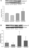
Arginase II is increased in hypoxia, and this hypoxia-induced increase in protein expression was attenuated by the EGFR inhibitor AG-1478. A: hypoxia has no effect on arginase I protein levels. Top: representative immunoblot from hPMVEC incubated with vehicle or 30 nM AG-1478 and placed in normoxia and hypoxia for 24 h. Bottom: graph of the mean densitometry data that were obtained from arginase I Western blotting analysis. B: hypoxia increased arginase II protein levels. Top: representative arginase II immunoblotting. Bottom: graph of the mean densitometry data obtained from arginase II immunoblot. *Vehicle-treated hypoxia different from vehicle-treated normoxia, P < 0.05. #AG-1478-treated cells significantly different from vehicle-treated cells in same condition, P < 0.05.
Fig. 7.
Fig. 7.
Urea production is increased in hypoxia, and this hypoxia-induced increase was prevented by the EGFR inhibitor AG-1478. Medium was collected from cells treated with AG-1478 (30 nM) or vehicle (n = 6 for each sample) and incubated in hypoxia or normoxia for 24 h. *Vehicle-treated cells in hypoxia different from normoxic vehicle-treated cells, P < 0.05. #AG-1478-treated cells different from vehicle-treated cells for the same condition, P < 0.05.
Fig. 8.
Fig. 8.
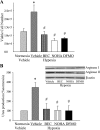
Arginase inhibition prevented hypoxia-induced cellular proliferation. A: proliferation assays from hypoxic hPMVECs treated with the arginase inhibitors α-difluoromethylornithine (DFMO; 100 mM), S-(2-boronoethyl)-l-cysteine (BEC; 100 μM), Nω-hydroxy-arginine (NOHA; 100 μM), or vehicle for 48 h. The number of viable cells was counted using Trypan blue exclusion. B: inhibition of arginase prevents hypoxia-induced urea production. Culture media were collected from hypoxic hPMVECs that were treated with vehicle, DFMO, BEC, or NOHA for 48 h. Inset: representative immunoblots for arginase I, arginase II, and β-actin to confirm that the arginase inhibitors had no substantial effect on arginase protein levels. *Hypoxia vehicle treated different from vehicle-treated normoxia, P < 0.05. #Arginase inhibitor different from vehicle-treated hypoxia, P < 0.05.
Fig. 9.
Fig. 9.
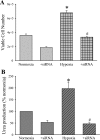
EGFR small interfering RNA (siRNA) prevented hypoxia-induced proliferation and urea production. A: hPMVEC were transfected with either vehicle, a scramble siRNA, or EGFR siRNA (Dharmacon) for 24 h. The hPMVEC were then washed, EGM placed on them, and the hPMVEC were allowed to recover in normoxia for 24 h. The hPMVEC were washed again and seeded at 200,000 cells per well on a six-well plate and placed in either normoxia or hypoxia for 24 h. The number of viable cells was then counted using Trypan blue exclusion. The vehicle-treated and scramble siRNA-treated hPMVEC were not different in either normoxia or hypoxia. B: medium was collected from hPMVEC treated with EGFR siRNA or vehicle (n = 6 for each sample) as above and assayed for urea production. The urea production was normalized to protein content of each plate and expressed as a percentage of normoxia. *Hypoxia vehicle treated different from vehicle-treated normoxia, P < 0.05. #+siRNA different from vehicle treated for the same condition, P < 0.05.
Fig. 10.
Fig. 10.
Proposed model for hypoxia-induced cellular proliferation via EGFR activation, with arginase as a necessary intermediate step. In our model, hypoxia results in EGFR activation, which leads to the induction of arginase II, which is required for EGFR-dependent, hypoxia-induced cellular proliferation. Pharmacological (AG-1478) or siRNA-mediated inhibition of EGFR expression and activation prevented both hypoxia-induced arginase II induction and cellular proliferation. Pharmacological inhibition of arginase activity (BEC, NOHA, and DFMO) prevented hypoxia-induced cellular proliferation.

Similar articles

Cited by

References

    1. Albina J, Mahoney E, Daley J, Reichner JS. Macrophage arginase regulation by CCAAT/enhancer-binding protein β. Shock 23: 168–172, 2005 - PubMed
    1. Bachetti T, Comini L, Francolini G, Bastianon D, Valetti B, Cadei M, Grigolato P, Ferrari R. Arginase pathway in human endothelial cells in pathophysiological conditions. J Mol Cell Cardiol 37: 515–523, 2004 - PubMed
    1. Baldwin BR, Timchenko NA, Zahnow CA. Epidermal growth factor receptor stimulation activates the RNA binding protein CUG-BP1 and increases expression of C/EBPβ-LIP in mammary epithelial cells. Mol Cell Biol 24: 3682–3691, 2004 - PMC - PubMed
    1. Banks M, Gerasimovskaya E, Tucker D, Stenmark K. Egr-1 antisense oligonucleotides inhibit hypoxia-induced proliferation of pulmonary artery adventitial fibroblasts. J Appl Physiol 98: 732–738, 2005 - PubMed
    1. Bivalacqua TJ, Burnett AL, Hellstrom WJ, Champion HC. Overexpression of arginase in the aged mouse penis impairs erectile function and decreases eNOS activity: influence of in vivo gene therapy of anti-arginase. Am J Physiol Heart Circ Physiol 292: H1340–H1351, 2007 - PubMed

Publication types

MeSH terms