. 2010 Mar 1;24(5):443-54.
doi: 10.1101/gad.1893210. Epub 2010 Feb 5.

The DMM complex prevents spreading of DNA methylation from transposons to nearby genes in Neurospora crassa

Affiliations

The DMM complex prevents spreading of DNA methylation from transposons to nearby genes in Neurospora crassa

Shinji Honda et al. Genes Dev. .

Abstract

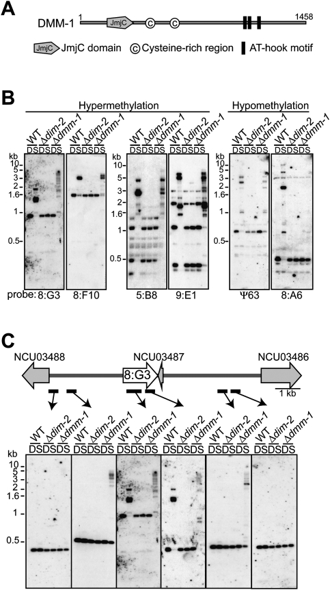
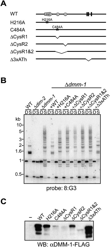
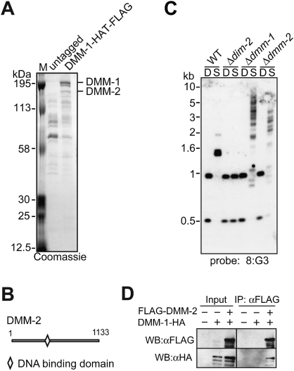
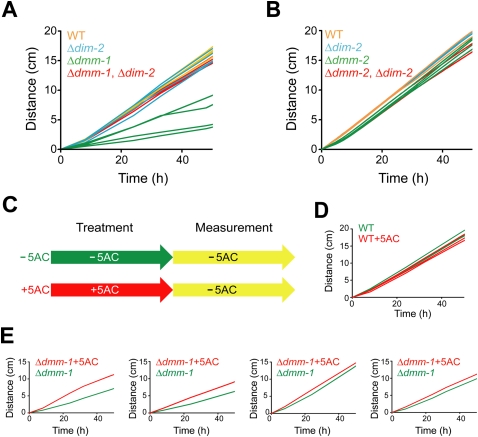
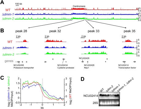
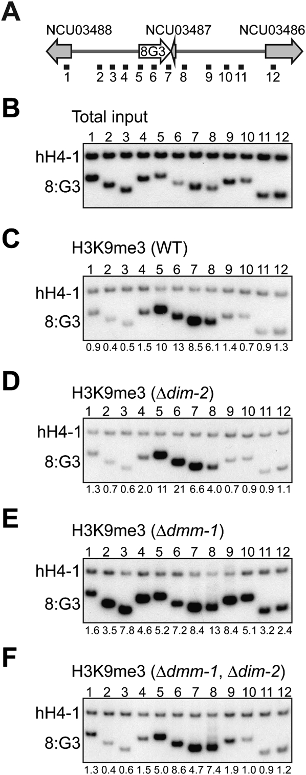
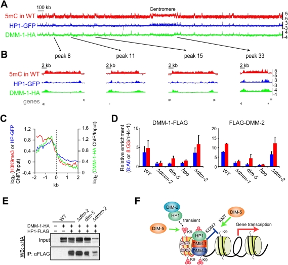
Transposable elements are common in genomes and must be controlled. Many organisms use DNA methylation to silence such selfish DNA, but the mechanisms that restrict the methylation to appropriate regions are largely unknown. We identified a JmjC domain protein in Neurospora, DNA METHYLATION MODULATOR-1 (DMM-1), that prevents aberrant spreading of DNA and histone H3K9 methylation from inactivated transposons into nearby genes. Mutation of a conserved residue within the JmjC Fe(II)-binding site abolished dmm-1 function, as did mutations in conserved cysteine-rich domains. Mutants defective only in dmm-1 mutants grow poorly, but growth is restored by reduction or elimination of DNA methylation using the drug 5-azacytosine or by mutation of the DNA methyltransferase gene dim-2. DMM-1 relies on an associated protein, DMM-2, which bears a DNA-binding motif, for localization and proper function. HP1 is required to recruit the DMM complex to the edges of methylated regions.


Figures

Figure 1.
Figure 1.
Mutants lacking the JmjC domain protein DMM-1 show aberrant DNA methylation. (A) Schematic representation of DMM-1 with predicted domains and amino acid coordinates indicated. (B) DNA from a Δdmm-1 strain is hypermethylated at 8:G3, 8:F10, 5:B8, and 9:E1, and hypomethylated at 8:A6 and Ψ63. Genomic DNA of a wild-type strain (WT), a DNA methyltransferase mutant (Δdim-2), and the dmm-1 mutant were digested with 5mC-sensitive Sau3AI (S) or its 5mC-insensitive isoschizomer, DpnII (D), and used for Southern hybridizations with the indicated probes for representative methylated regions (Selker et al. 2003). (C) Spreading of DNA methylation from the methylated 8:G3 region in the dmm-1 mutant. The genomic region is shown schematically above the results of a Southern hybridization probed for 8:G3 (white arrow) and the neighboring regions (probes indicated by bars), which include a RIP'd pseudogene (NCU03487) and two predicted genes (NCU03486 and NCU03488) (gray arrow).
Figure 2.
Figure 2.
The DMM-1 JmjC domain and cysteine-rich (CysR) regions, but not AT hook motifs, are essential for normal DNA methylation. (A) Diagram of the DMM-1 point mutations and deletion mutations tested. His 216 is predicted to be critical for Fe(II) binding, and Cys 484 is conserved among fungal DMM-1 homologs (see Supplemental Figs. 3B, 4). (B) Effects of dmm-1 mutations on DNA methylation in 8:G3 region. DNA isolated from indicated strains was analyzed by Southern blot as described in Figure 1B. (C) Expression of dmm-1 mutants was assessed by Western blotting with antibodies against Flag.
Figure 3.
Figure 3.
DMM-1 is associated with DMM-2. (A) Purification and mass spectrometric analyses of proteins associated with DMM-1. Extracts from strains containing untagged or HAT-Flag-tagged DMM-1 were subjected to a two-step purification, separated by SDS-PAGE, and visualized by Coomassie blue staining, and proteins from individual bands were digested with trypsin and analyzed by tandem mass spectrometry. (B) Schematic representation of the predicted 1133-amino-acid DMM-2 protein showing the position of a Zn(II)2Cys6 putative DNA-binding domain. (C) A mutant lacking DMM-2 shows hypermethylation at 8:G3 region. DNA isolated from the indicated strains was analyzed by Southern blot as described in Figure 1B. (D) DMM-2 associates with DMM-1 in vivo. Extracts from strains with (+) or without (−) the Flag-tagged dmm-2 gene and/or the HA-tagged dmm-1 gene were immunoprecipitated with anti-Flag antibodies. Input and immunoprecipitation samples were fractionated and analyzed by Western blotting with the indicated antibodies.
Figure 4.
Figure 4.
The growth defect in Δdmm-1 strains is relieved by loss of DNA methylation. (A) Growth rates at 32°C on solidified Vogel's medium in “race tubes” are shown for four wild-type strains (WT; orange), four Δdim-2 mutants (blue), four Δdmm-1 mutants (green), and four Δdmm-1, Δdim-2 double mutants (red). (B) Δdmm-2 strains do not show a growth defect under the same conditions. Four Δdmm-2 mutants (green) and four Δdmm-2, Δdim-2 double mutants (red) along with control strains as in A. (C) Schematic diagram of 5AC treatments. Strains were grown on solidified Vogel's medium for 3 d, followed by liquid culture for 5 h with (+) or without (−) 5AC, and linear growth rates were determined in race tubes containing solidified Vogel's medium without 5AC. (D,E) Growth rates are shown for four wild-type strains (WT) and four Δdmm-1 strains pretreated with (red) or without (green) 5AC.
Figure 5.
Figure 5.
DNA methylation spreads into neighboring genes in Δdmm-1 but not Δdmm-2 strains. (A) DNA methylation profile of wild-type (WT), Δdmm-1, and Δdmm-2 strains across entire chromosome VII. Methylated DNA were immunoprecipitated (MeDIP) from DNA of wild-type (red), Δdmm-1 (blue), and Δdmm-2 (green) strains and used to probe ∼40,000 oligonucleotide sequences on an Agilent slide (Lewis et al. 2008). Results are shown as log2[IP/input] values (Y-axis). A scale bar is shown, and the centromere region is identified. (B) Data for four representative methylated regions of chromosome VII (positions indicated by arrows) are shown magnified ∼50-fold. (Bottom) The positions of predicted genes are shown in gray, and those that are methylated in Δdmm-1 but not Δdmm-2 are indicated. (C) Averaged levels of DNA methylation across the ∼80 edges of methylated regions on chromosome VII in wild-type (WT, red), Δdmm-1 (blue), and Δdmm-2 (green) strains. (D) Down-regulation of NCU02414 gene in a Δdmm-1 strain is relieved by loss of DNA methylation. Total RNA of the indicated strains were isolated and used for Northern hybridization with an NCU02414 probe. 28S rRNA was visualized by ethidium bromide (EtBr) staining.
Figure 6.
Figure 6.
H3K9me3 spreads in Δdmm-1 mutants but not in Δdmm-1, Δdim-2 double mutants. (A) Schematic representation of the 8:G3 region (see Fig. 1B). The numbered horizontal black bars indicate the regions tested on total input DNA (example for wild type shown in B) or by ChIP using antibodies against H3K9me3 (C–F) using chromatin from the indicated strains. A euchromatic gene lacking DNA methylation (hH4-1) was used as an internal control. The ratios of intensities measured for hH4-1 and the indicated probe were normalized to the ratios obtained without immunoprecipitation (total input) and are presented below the lanes. ChIP experiments were repeated at least twice with equivalent results (data not shown).
Figure 7.
Figure 7.
DMM-1 is localized preferentially at edges of methylated regions via associations with HP1 and DMM-2. (A) MeDIP (red) and ChIP–chip analyses with antibodies against HP1-GFP (blue) and DMM-1-HA (green) in otherwise wild-type (WT) strains are shown as log2[IP/input] values (Y-axis). (B) Data for four representative methylated regions of chromosome VII (positions indicated by arrows) are shown magnified ∼50-fold. (Bottom) The positions of predicted genes are shown in gray. (C) Average enrichment of H3K9me3 (red), HP1-GFP (blue), and DMM-1-HA (green) on the 80 edges of methylated regions of chromosome VII. (D) Conventional ChIP analyses with antibodies against DMM-1-Flag or Flag-DMM-2 in indicated strains at two methylated regions: 8:A6 and the center of 8:G3 (segment 6 in Fig. 6A). Data from duplicate ChIP experiments (Supplemental Fig. 22B) were averaged and are presented graphically with error bars to indicate variation. (E) DMM-1 association with HP1 is independent of DIM-2, DIM-5, and DMM-2. Extracts from strains with (+) or without (−) the HA-tagged dmm-1 gene and/or the Flag-tagged hpo gene in the indicated strains were immunoprecipitated with anti-Flag antibodies. Input and immunoprecipitation samples were fractionated and analyzed by Western blotting with anti-HA antibodies. (F) Model for the involvement of HP1/DMM-1/2 complex in normal distribution of DNA methylation within transposon relics. (Red lines) RIP'd/A:T-rich DNA; (black lines) unRIP'd DNA; (small red circles) methyl groups; (mC) methyl-cytosine; (KDM) lysine demethylase.

Similar articles

Cited by

References

    1. Agger K, Christensen J, Cloos PA, Helin K. The emerging functions of histone demethylases. Curr Opin Genet Dev. 2008;18:159–168. - PubMed
    1. Ayoub N, Noma K, Isaac S, Kahan T, Grewal SI, Cohen A. A novel jmjC domain protein modulates heterochromatization in fission yeast. Mol Cell Biol. 2003;23:4356–4370. - PMC - PubMed
    1. Cambareri EB, Foss HM, Rountree MR, Selker EU, Kinsey JA. Epigenetic control of a transposon-inactivated gene in Neurospora is dependent on DNA methylation. Genetics. 1996;143:137–146. - PMC - PubMed
    1. Chan SW, Henderson IR, Jacobsen SE. Gardening the genome: DNA methylation in Arabidopsis thaliana. Nat Rev Genet. 2005;6:351–360. - PubMed
    1. Colot HV, Park G, Turner GE, Ringelberg C, Crew CM, Litvinkova L, Weiss RL, Borkovich KA, Dunlap JC. A high-throughput gene knockout procedure for Neurospora reveals functions for multiple transcription factors. Proc Natl Acad Sci. 2006;103:10352–10357. - PMC - PubMed

Publication types

MeSH terms

Substances