

Activated alleles of the Schizosaccharomyces pombe gpa2+ Galpha gene identify residues involved in GDP-GTP exchange
- PMID: 20139237
- PMCID: PMC2863420
- DOI: 10.1128/EC.00010-10
Activated alleles of the Schizosaccharomyces pombe gpa2+ Galpha gene identify residues involved in GDP-GTP exchange
Abstract
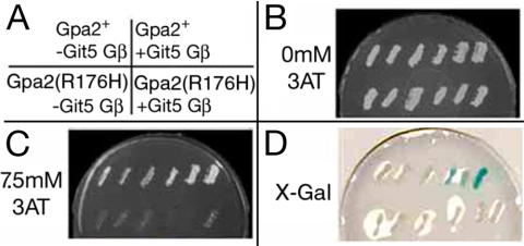
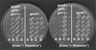
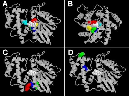
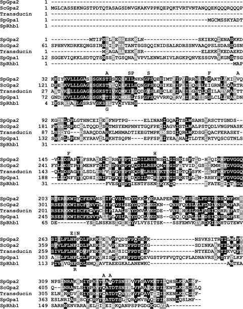
The Schizosaccharomyces pombe glucose/cyclic AMP (cAMP) signaling pathway includes the Gpa2-Git5-Git11 heterotrimeric G protein, whose Gpa2 Galpha subunit directly binds to and activates adenylate cyclase in response to signaling from the Git3 G protein-coupled receptor. To study intrinsic and extrinsic regulation of Gpa2, we developed a plasmid-based screen to identify mutationally activated gpa2 alleles that bypass the loss of the Git5-Git11 Gbetagamma dimer to repress transcription of the glucose-regulated fbp1(+) gene. Fifteen independently isolated mutations alter 11 different Gpa2 residues, with all but one conferring a receptor-independent activated phenotype upon integration into the gpa2(+) chromosomal locus. Biochemical characterization of three activated Gpa2 proteins demonstrated an increased GDP-GTP exchange rate that would explain the mechanism of activation. Interestingly, the amino acid altered in the Gpa2(V90A) exchange rate mutant protein is in a region of Gpa2 with no obvious role in Galpha function, thus extending our understanding of Galpha protein structure-function relationships.
Figures
Similar articles
-
Glucose monitoring in fission yeast via the Gpa2 galpha, the git5 Gbeta and the git3 putative glucose receptor.Genetics. 2000 Oct;156(2):513-21. doi: 10.1093/genetics/156.2.513. Genetics. 2000. PMID: 11014802 Free PMC article.
-
The git5 Gbeta and git11 Ggamma form an atypical Gbetagamma dimer acting in the fission yeast glucose/cAMP pathway.Genetics. 2001 Mar;157(3):1159-68. doi: 10.1093/genetics/157.3.1159. Genetics. 2001. PMID: 11238401 Free PMC article.
-
Sck1 negatively regulates Gpa2-mediated glucose signaling in Schizosaccharomyces pombe.Eukaryot Cell. 2014 Feb;13(2):202-8. doi: 10.1128/EC.00277-13. Epub 2013 Dec 2. Eukaryot Cell. 2014. PMID: 24297439 Free PMC article.
-
Glucose sensing via the protein kinase A pathway in Schizosaccharomyces pombe.Biochem Soc Trans. 2005 Feb;33(Pt 1):257-60. doi: 10.1042/BST0330257. Biochem Soc Trans. 2005. PMID: 15667320 Free PMC article. Review.
-
Heterotrimeric G Proteins in Plants: Canonical and Atypical Gα Subunits.Int J Mol Sci. 2021 Oct 31;22(21):11841. doi: 10.3390/ijms222111841. Int J Mol Sci. 2021. PMID: 34769272 Free PMC article. Review.
Cited by
-
Rapid, efficient and precise allele replacement in the fission yeast Schizosaccharomyces pombe.Curr Genet. 2014 May;60(2):109-19. doi: 10.1007/s00294-013-0406-x. Epub 2013 Sep 12. Curr Genet. 2014. PMID: 24026504 Free PMC article.
-
An Ancient Yeast for Young Geneticists: A Primer on the Schizosaccharomyces pombe Model System.Genetics. 2015 Oct;201(2):403-23. doi: 10.1534/genetics.115.181503. Genetics. 2015. PMID: 26447128 Free PMC article. Review.
-
A genomewide screen in Schizosaccharomyces pombe for genes affecting the sensitivity of antifungal drugs that target ergosterol biosynthesis.Antimicrob Agents Chemother. 2012 Apr;56(4):1949-59. doi: 10.1128/AAC.05126-11. Epub 2012 Jan 17. Antimicrob Agents Chemother. 2012. PMID: 22252817 Free PMC article.
-
Use of a ura5+-lys7+ cassette to construct unmarked gene knock-ins in Schizosaccharomyces pombe.Curr Genet. 2012 Feb;58(1):59-64. doi: 10.1007/s00294-011-0360-4. Epub 2011 Dec 25. Curr Genet. 2012. PMID: 22198627
-
A yeast-based chemical screen identifies a PDE inhibitor that elevates steroidogenesis in mouse Leydig cells via PDE8 and PDE4 inhibition.PLoS One. 2013 Aug 14;8(8):e71279. doi: 10.1371/journal.pone.0071279. eCollection 2013. PLoS One. 2013. PMID: 23967182 Free PMC article.
References
-
- Apanovitch D. M., Iiri T., Karasawa T., Bourne H. R., Dohlman H. G. 1998. Second site suppressor mutations of a GTPase-deficient G-protein alpha-subunit. Selective inhibition of Gbeta gamma-mediated signaling. J. Biol. Chem. 273:28597–28602 - PubMed
-
- Bähler J., Wu J. Q., Longtine M. S., Shah N. G., McKenzie A., III, Steever A. B., Wach A., Philippsen P., Pringle J. R. 1998. Heterologous modules for efficient and versatile PCR-based gene targeting in Schizosaccharomyces pombe. Yeast 14:943–951 - PubMed
-
- Basi G., Schmid E., Maundrell K. 1993. TATA box mutations in the Schizosaccharomyces pombe nmt1 promoter affect transcription efficiency but not the transcription start point or thiamine repressibility. Gene 123:131–136 - PubMed
-
- Chidiac P., Ross E. M. 1999. Phospholipase C-beta1 directly accelerates GTP hydrolysis by Galphaq and acceleration is inhibited by Gbeta gamma subunits. J. Biol. Chem. 274:19639–19643 - PubMed
-
- Coleman D. E., Lee E., Mixon M. B., Linder M. E., Berghuis A. M., Gilman A. G., Sprang S. R. 1994. Crystallization and preliminary crystallographic studies of Gi alpha 1 and mutants of Gi alpha 1 in the GTP and GDP-bound states. J. Mol. Biol. 238:630–634 - PubMed
Publication types
MeSH terms
Substances
Grants and funding
LinkOut - more resources
Full Text Sources
Molecular Biology Databases
Miscellaneous
