

Odontogenic myxofibroma synchronous with primary angiosarcoma of the spleen
- PMID: 20139242
- PMCID: PMC3487256
- DOI: 10.1259/bjr/14078580
Odontogenic myxofibroma synchronous with primary angiosarcoma of the spleen
Abstract
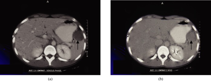
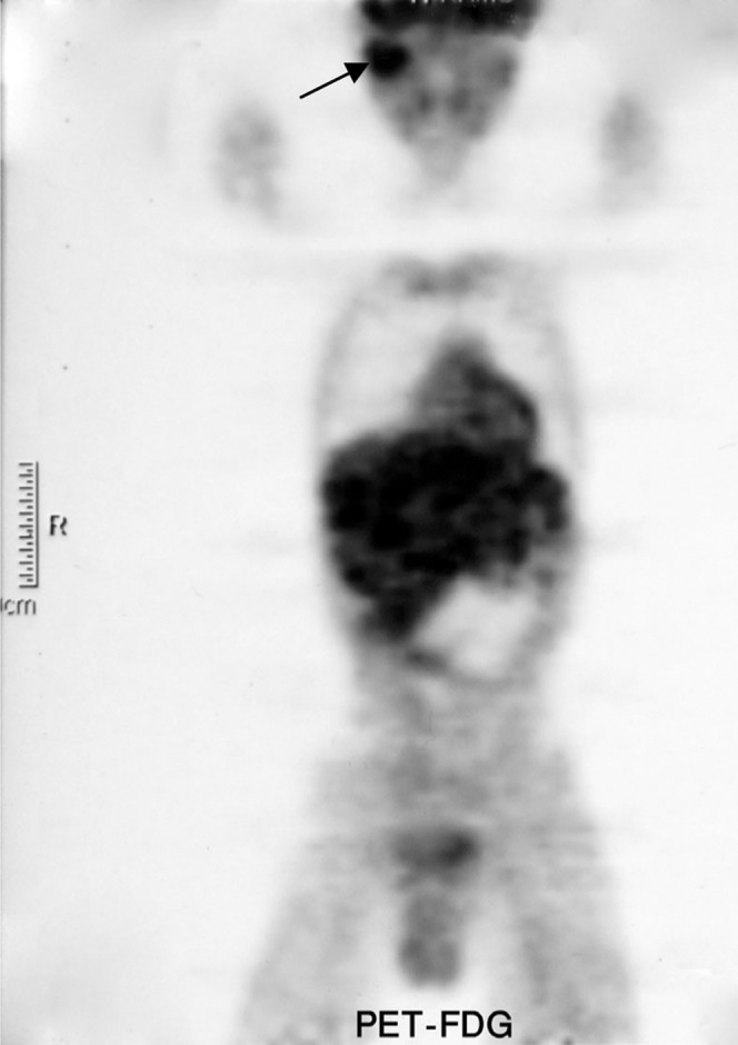
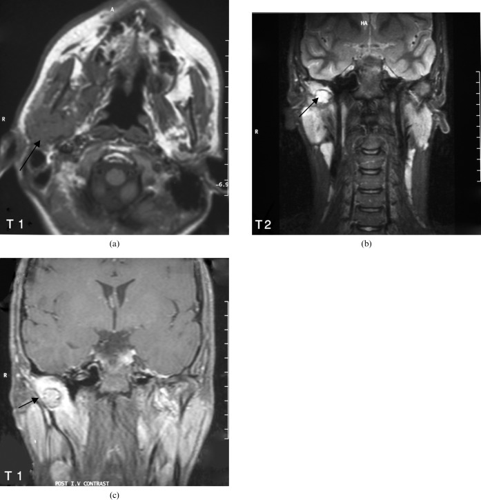
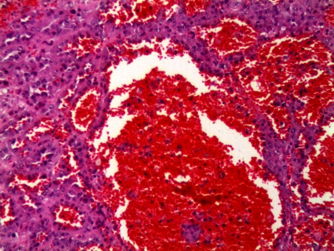
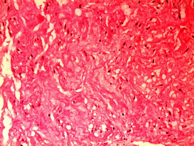
Odontogenic myxofibroma of the temporomandibular joint (TMJ) is a rare tumour; moreover, primary splenic angiosarcoma (PAS) in paediatric patients is extremely rare. We report on a 15-year-old boy who presented with right TMJ swelling and subsequently developed epigastric and right upper quadrant pain. The TMJ swelling proved to be odontogenic myxofibroma and the abdominal pain was a result of primary splenic angiosarcoma with hepatic metastasis. We report for the first time the synchronous presentation of PAS and odontogenic myxofibroma in a paediatric patient, and we describe the radiological features along with the histological diagnosis and clinical outcome. Uptake in (18)F-2-fluoro-2-deoxy-D-glucose positron emission tomography is also described for the first time for both these tumours.
Figures
References
-
- Hsu J, Ueng S, Hwang T, Chen H, Jan Y, Chen M. Primary angiosarcoma of the spleen in a child with long-term survival. Pediatr Surg Int 2007;23:807–10 - PubMed
-
- Vrchliotis T, Bennett W, Vaswani K, Niemann T, Bova J. Primary angiosarcoma of the spleen- CT, MR, and sonographic characteristics: report of two cases. Abdom Imaging 2000;25:283–5 - PubMed
-
- Hoed I, Granzen B, Aronson D, Pauwels P, Kraker J, Heurn L. Metastasized angiosarcoma of the spleen in a 2-year-old girl. Pediatr Hematol Oncol 2005; 22:5,387–90. - PubMed
-
- Thompson W, Aguilera N, Gorospe L, Abbott R. Angiosarcoma of the spleen: Imaging characteristics in 12 patients. Radiology 2005;235:106–15 - PubMed
Publication types
MeSH terms
LinkOut - more resources
Full Text Sources
Medical
