

Case Reports
doi: 10.1259/bjr/50945216.
Cryptococcal choroid plexitis: rare imaging findings of central nervous system cryptococcal infection in an immunocompetent individual
Affiliations
- PMID: 20139243
- PMCID: PMC3487264
- DOI: 10.1259/bjr/50945216
Item in Clipboard
Case Reports
Cryptococcal choroid plexitis: rare imaging findings of central nervous system cryptococcal infection in an immunocompetent individual
Br J Radiol.
2010 Jan.
Abstract
Central nervous system (CNS) cryptococcosis is a common opportunistic fungal infection in immunocompromised patients, and the imaging findings differ from those in immunocompetent patients. Here, we present the imaging findings in an immunocompetent woman of a rare case of central nervous system cryptococcal choroid plexitis with trapped temporal horns, enlarged enhancing bilateral choroid plexuses and multiple intraventricular choroid plexus cysts.
Figures
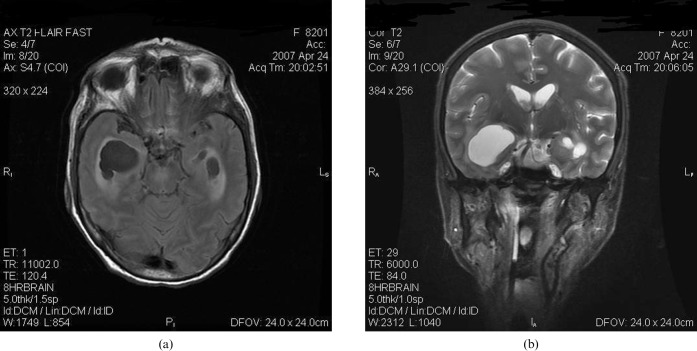
(a) Axial T2 fluid-attenuated inversion-recovery and (b) coronal T2 weighted images show asymmetrically dilated and trapped temporal horns with periventricular extravasation of cerebrospinal fluid. Prominent perivascular spaces with adjacent oedema can be noted in the left basal ganglia.
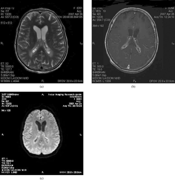
(a) Axial T2 weighted image at the level of the lateral ventricular trigones reveals multiple bilateral intraventricular choroid plexus cysts and adjacent periventricular white matter oedema. (c) Contrast-enhanced axial T1 weighted image at the same level shows multilocular rim enhancement in both choroid plexuses. (c) Diffusion-weighted image (b-value _ 1000) shows no significant restriction of diffusion.
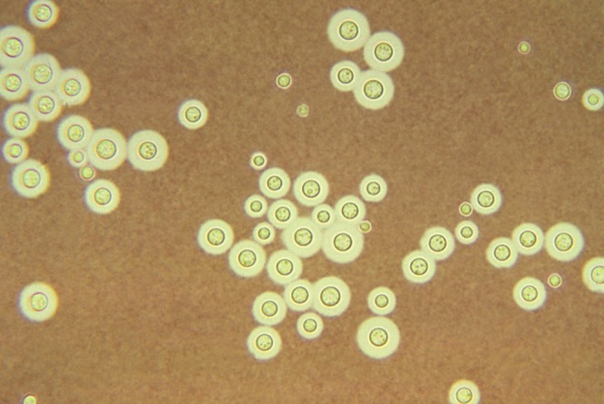
A photomicrograph of light Indian ink preparation of cerebrospinal fluid shows the typical encapsulated yeast cells.
References
-
- Kuroki M, Phichaichumpon C, Yasuoka A, Chiranairadul P, Chosa T, Sirinirund P, et al. Enviornmental isolation of Cryptococcus neoformans from an epidemic region of HIV associated cryptococcus in Thailand. Yeast 2004;21:809–12 - PubMed
-
- Igel HJ, Bolande RP. Humoral defense mechanisms in cryptococcosis; substances in normal human serum, saliva and CSF affecting growth of Cryptococcus neoformans. J Infect Dis 1966:116–75 - PubMed
-
- Andreula CF, Burdi N, Carella A. CNS cryptococosis in AIDS. Spectrum of MR findings. J Comput Tomogr 1993;17:438–41 - PubMed
