Case Reports
. 2010 Jan;83(985):e18-21.
doi: 10.1259/bjr/66268641.

Intracranial transthecal subarachnoid fat emboli and subarachnoid haemorrhage arising from a sacral fracture and dural tear

Affiliations
Case Reports

Intracranial transthecal subarachnoid fat emboli and subarachnoid haemorrhage arising from a sacral fracture and dural tear

J K H Woo et al. Br J Radiol. 2010 Jan.

Abstract

We present the case of a 28-year-old man with an unusual aetiology of lipid-dense material in the subarachnoid space. CT of the head at presentation was normal. MRI of the spine revealed a defect in the dura at L5/S1, with avulsed left L5 and S1 nerve roots. Haematoma and marrow fat were observed in close relation to the dural tear adjacent to the sacral fracture. Head CT and MRI subsequently demonstrated new lipid-dense material and haemorrhage in the subarachnoid space after sacral instrumentation, presumably owing to transthecal displacement of fatty marrow.


Figures

Figure 1
Figure 1
Axial non-contrast CT images with wide windows at the level of the prepontine cistern at (a) presentation and at (b) 3 days post-trauma. (a) Normal cerebrospinal fluid is seen in the prepontine cistern (arrow). (b) Globules and linear strands of fat (−124 HU) are seen in the prepontine cistern (arrowheads).
Figure 2
Figure 2
Axial non-contrast T1 weighted MR images of the brain three days post-trauma. (a) Hyperintense non-dependent fat globules are seen in the prepontine cistern (arrow). (b) Tiny hyperintense fat globules are present in the anterior aspect of the frontal horns (arrows) with haemorrhage in the trigones (arrowheads) bilaterally.
Figure 3
Figure 3
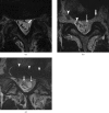
Axial non-contrast T2 weighted MR images of the spine three days post-trauma. (a) A focal defect is shown in the left anterolateral aspect of the thecal sac at the level of the L5 superior endplate (arrow). (b) An avulsed left L5 nerve root (arrow), absence of the left S1 nerve root and epidural hematoma (squiggly arrow) at the level of the L5/S1 disc are also indicated, as are the right L5 and S1 nerve roots (arrowheads). (c) A dural tear with blood and fat extending into thecal sac (arrow) is seen at the level of the L5/S1 disc. The image also shows a small pseudomeningocele (squiggly arrow) and an iliosacral screw (arrowheads).

References

    1. Simon AD, Ulmer JL, Strottmann JM. Contrast-enhanced MR imaging of cerebral fat embolism: case report and review of the literature. AJNR AM J Neuroradiol 2003;24:97–101 - PMC - PubMed
    1. Lee TC, Bartlett ES, Fox AJ, Symons SP. The hypodense artery sign. AJNR AM J Neuroradiol 2005;26:2027–29 - PMC - PubMed
    1. Smith AS, Benson JE, Blaser SI, Mizushima A, Tarr RW, Bellon EM. Diagnosis of ruptured intracranial dermoid cyst: value MR over CT. AJNR AM J Neuroradiol 1991;12:175–80 - PMC - PubMed
    1. Bhangoo RS, Tammam A, Crockard HA. MRI detection of spontaneous rupture of a well differentiated pineal teratoma. Acta Neurochir (Wien) 1997;139:891–2 - PubMed
    1. Harrison RL, Abernethy LJ. Asymptomatic intraventricular lipid leak from a primary pineal teratoma. Pediatr Radiol 2001;31:129–31 - PubMed

Publication types

MeSH terms