MR spectroscopy and MR perfusion character of cerebral sparganosis: a case report
- PMID: 20139254
- PMCID: PMC3473539
- DOI: 10.1259/bjr/73038348
MR spectroscopy and MR perfusion character of cerebral sparganosis: a case report
Abstract
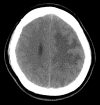
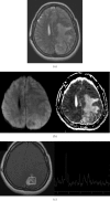
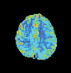
The authors report the case of a 46-year-old woman with cerebral sparganosis resulting from infection with a larva of Spirometra. Computed tomography and magnetic resonance imaging revealed a mass lesion with prominent perifocal oedema in the left parietal lobe. Advanced imaging pulse sequences, including MR spectroscopy and MR perfusion, were performed. During surgery for the removal of a granuloma, the parasite was discovered and excised. Following treatment, the patient's neurological deficits markedly improved.
Figures


Comment in
-
Cerebral sparganosis.Br J Radiol. 2010 Sep;83(993):807. doi: 10.1259/bjr/22825470. Epub 2010 Aug 17. Br J Radiol. 2010. PMID: 20716651 Free PMC article. No abstract available.
References
-
- Cummings TJ, Madden JF, Gray L, Friedman AH, McLendon RE. Parasitic lesion of the insula suggesting cerebral sparganosis: case report. Neuroradiology 2000;42:206–8 - PubMed
-
- Tsai MD, Chang CN, Ho YS, Wang AD. Cerebral sparganosis diagnosed and treated with stereotactic techniques. Report of two cases. J Neurosurg 1993;78:129–32 - PubMed
-
- Chang KH, Cho SY, Chi JG, Kim WS, Han MC, Kim CW, et al. Cerebral sparganosis: CT characteristics. Radiology 1987;165:505–10 - PubMed
-
- Bo G, Xuejian W. Neuroimaging and pathological findings in a child with cerebral sparganosis. Case report. J Neurosurg 2006;105:470–2 - PubMed
Publication types
MeSH terms
LinkOut - more resources
Full Text Sources
Medical

