

Case Reports
doi: 10.1259/bjr/73038348.
MR spectroscopy and MR perfusion character of cerebral sparganosis: a case report
Affiliations
- PMID: 20139254
- PMCID: PMC3473539
- DOI: 10.1259/bjr/73038348
Item in Clipboard
Case Reports
MR spectroscopy and MR perfusion character of cerebral sparganosis: a case report
Br J Radiol.
2010 Feb.
Abstract
The authors report the case of a 46-year-old woman with cerebral sparganosis resulting from infection with a larva of Spirometra. Computed tomography and magnetic resonance imaging revealed a mass lesion with prominent perifocal oedema in the left parietal lobe. Advanced imaging pulse sequences, including MR spectroscopy and MR perfusion, were performed. During surgery for the removal of a granuloma, the parasite was discovered and excised. Following treatment, the patient's neurological deficits markedly improved.
Figures
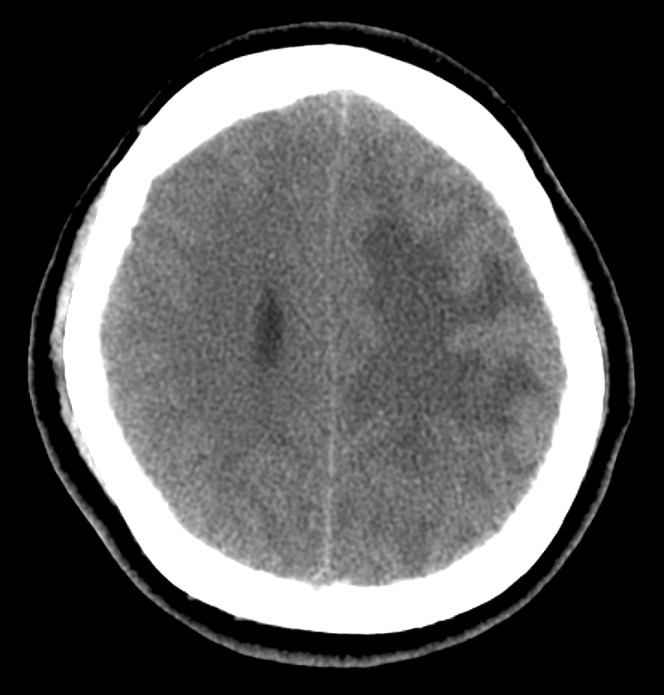
Pre-contrast CT scan shows a patchy area of hypoattenuation in the white matter of the left parietal lobe with prominent mass effect.
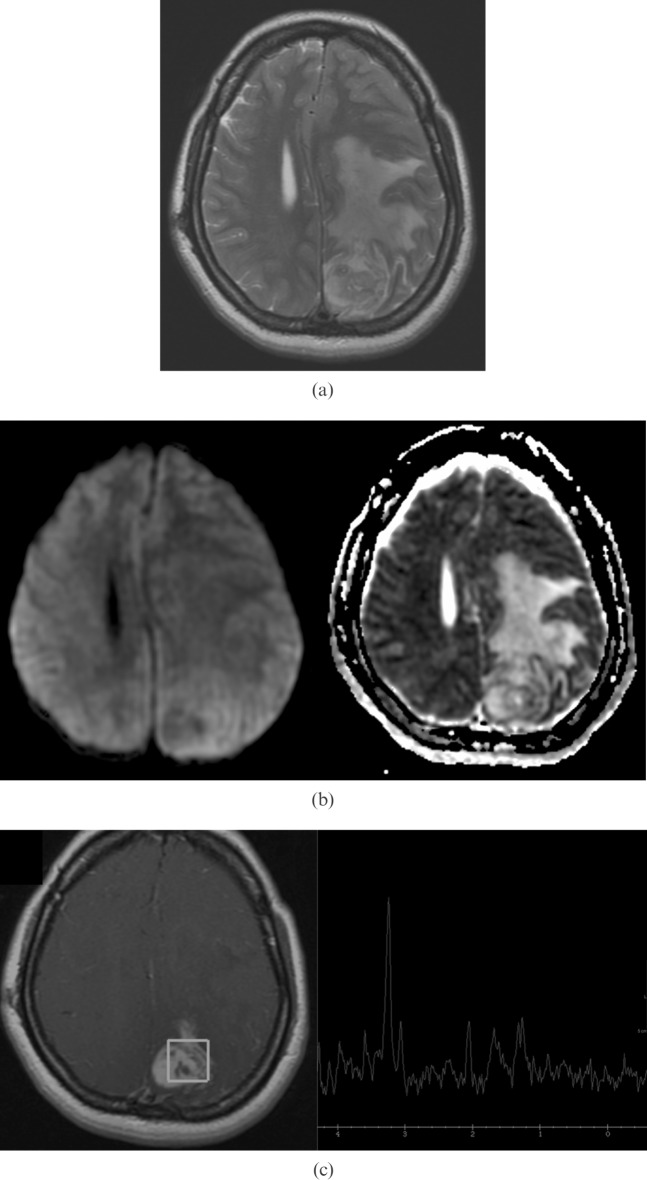
(a) Axial T2 weighted MRI shows a mixed iso-to-hyposignal lesion in the left high parietal lobe with extensive surrounding oedema. (b) Diffusion weighted imaging (left) and apparent diffusion coefficient mapping imaging (right), the lesion shows increased signal intensity. (c) MR spectroscopy (TE _ 144 ms) reveals an increased choline peak and decreased creatine and N-acetylaspartate peaks. There is a lipid or lactate doubling peak at 1.3 ppm. An elevated peak between 1.4 to 1.8 ppm is suspected to be alanine.
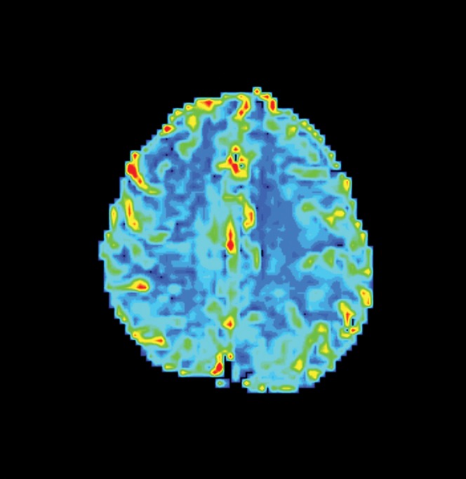
MR perfusion imaging shows no apparent increased cerebral perfusion of the lesion and surrounding area.
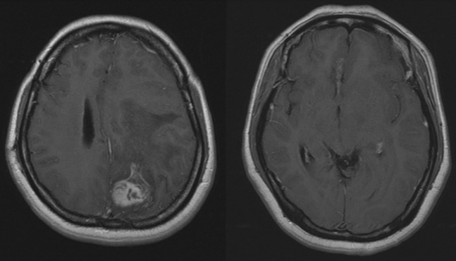
Post-contrast T1 weighted images show a heterogeneous enhanced parietal lobe lesion and a small rim-enhanced tunnel lesion in the gyrus rectus of the right frontal lobe.
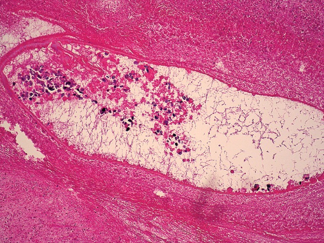
Photogram of a section of the lesion reveals an inflammatory granuloma with a dead parasite body. Inside the parasite, calcareous bodies are evident. Haematoxylin and eosin (H&E) stain.
Comment in
-
Cerebral sparganosis.Br J Radiol. 2010 Sep;83(993):807. doi: 10.1259/bjr/22825470. Epub 2010 Aug 17. Br J Radiol. 2010. PMID: 20716651 Free PMC article. No abstract available.
References
-
- Cummings TJ, Madden JF, Gray L, Friedman AH, McLendon RE. Parasitic lesion of the insula suggesting cerebral sparganosis: case report. Neuroradiology 2000;42:206–8 - PubMed
-
- Tsai MD, Chang CN, Ho YS, Wang AD. Cerebral sparganosis diagnosed and treated with stereotactic techniques. Report of two cases. J Neurosurg 1993;78:129–32 - PubMed
-
- Chang KH, Cho SY, Chi JG, Kim WS, Han MC, Kim CW, et al. Cerebral sparganosis: CT characteristics. Radiology 1987;165:505–10 - PubMed
-
- Bo G, Xuejian W. Neuroimaging and pathological findings in a child with cerebral sparganosis. Case report. J Neurosurg 2006;105:470–2 - PubMed
Publication types
MeSH terms
LinkOut - more resources
Full Text Sources
Medical
