

Case report. PET/CT appearance of desmoid tumour of the chest wall
- PMID: 20139256
- PMCID: PMC3473531
- DOI: 10.1259/bjr/18648939
Case report. PET/CT appearance of desmoid tumour of the chest wall
Abstract
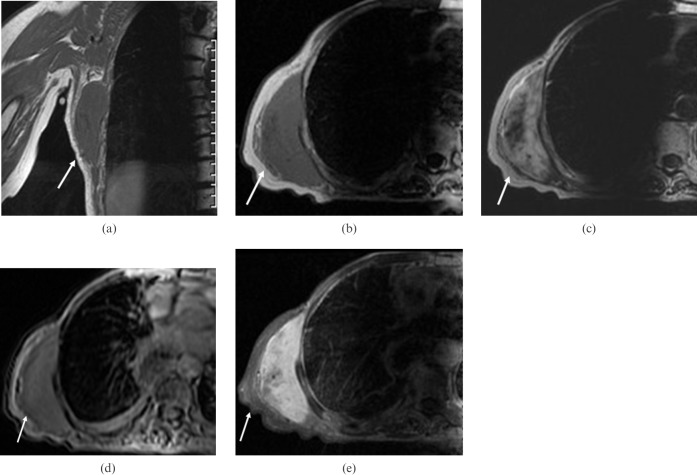
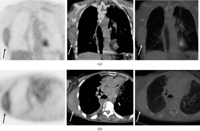
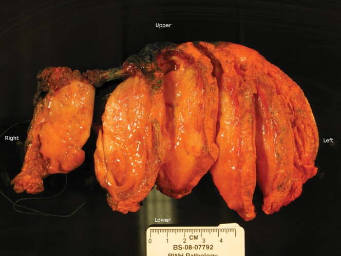
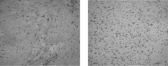
Desmoid tumours are rare, poorly circumscribed tumours that have a firm consistency and, although benign, have a remarkable tendency to infiltrate into surrounding structures. Extra-abdominal desmoid tumours involve mainly the extremities or the chest wall and are usually managed by wide radical resection. Moreover, desmoid tumours involving the chest wall are locally aggressive tumours with a high recurrence rate. We report a case of a pathologically proven desmoid tumour of the chest wall in a patient with a history of bilateral breast cancer and oesophageal cancer. We discuss the imaging appearances of this tumour on positron emission tomography combined with computed tomography (PET/CT) and magnetic resonance imaging.
Figures
Similar articles
-
Desmoid tumor of the chest wall in an elderly woman.Gen Thorac Cardiovasc Surg. 2009 Oct;57(10):554-7. doi: 10.1007/s11748-008-0404-y. Epub 2009 Oct 16. Gen Thorac Cardiovasc Surg. 2009. PMID: 19830521
-
[Desmoid tumor of the thoraco-abdominal wall characterized with 18F-fluorodeoxyglucose PET/ CT scan. Correlation with magnetic resonance and bone scintigraphy. Review of the literature].Rev Esp Med Nucl. 2009 Mar-Apr;28(2):70-3. doi: 10.1016/s0212-6982(09)70700-1. Rev Esp Med Nucl. 2009. PMID: 19406052 Review. Spanish.
-
Multicentric extra-abdominal desmoid tumours.Br J Plast Surg. 2004 Jun;57(4):362-5. doi: 10.1016/j.bjps.2004.02.014. Br J Plast Surg. 2004. PMID: 15145742 Review.
-
Desmoid-type chest wall fibromatosis. A six cases series.Orthop Traumatol Surg Res. 2011 Feb;97(1):102-7. doi: 10.1016/j.otsr.2010.09.017. Epub 2011 Jan 15. Orthop Traumatol Surg Res. 2011. PMID: 21239240
-
Musculoskeletal desmoid tumours: Diagnostic imaging appearances.J Med Imaging Radiat Oncol. 2015 Aug;59(4):461-467. doi: 10.1111/1754-9485.12318. Epub 2015 May 13. J Med Imaging Radiat Oncol. 2015. PMID: 25974678
Cited by
-
Mesenteric Fibromatosis Mimicking Metastasis: A Case Report and Review of Literature.J Gastrointest Cancer. 2012 Sep;43 Suppl 1:S73-6. doi: 10.1007/s12029-011-9298-5. J Gastrointest Cancer. 2012. PMID: 21710175 Review. No abstract available.
-
Fibromatosis with aggressive demeanor: Benign impersonator of malignancy.World J Nucl Med. 2020 Dec 12;20(1):121-124. doi: 10.4103/wjnm.WJNM_55_20. eCollection 2021 Jan-Mar. World J Nucl Med. 2020. PMID: 33850503 Free PMC article.
-
Fibromatosis arising from the pectoralis major muscle mimicking breast cancer.Radiol Case Rep. 2018 Sep 13;13(6):1174-1178. doi: 10.1016/j.radcr.2018.08.017. eCollection 2018 Dec. Radiol Case Rep. 2018. PMID: 30233754 Free PMC article.
-
Desmoid tumour (aggressive fibromatosis) of the colon mimics malignancy on dual time-point 18F-FDG PET/CT imaging.Br J Radiol. 2012 Feb;85(1010):e37-40. doi: 10.1259/bjr/43870228. Br J Radiol. 2012. PMID: 22308225 Free PMC article.
-
Imaging characteristics of spindle cell lipoma and its variants.Skeletal Radiol. 2014 May;43(5):591-7. doi: 10.1007/s00256-014-1834-5. Epub 2014 Feb 20. Skeletal Radiol. 2014. PMID: 24554024 Review.
References
-
- Dominguez-Malagon HR, Alfeiran-Ruiz A, Chavarria-Xicotencatl P, Duran-Hernandez MS. Clinical and cellular effects of colchicine in fibromatosis. Cancer 1992;69:2478–83 - PubMed
-
- Eubank WB, Mankoff DA. Evolving role of positron emission tomography in breast cancer imaging. Semin Nucl Med 2005;35:84–99 - PubMed
-
- Tateishi U, Yamaguchi U, Seki K, Terauchi T, Arai Y, Kim E. Bone and soft-tissue sarcoma: preoperative staging with fluorine 18 fluorodeoxyglucose PET/CT and conventional imaging. Radiology 2007;245:839–47 - PubMed
-
- Chapelier AR, Bacha EA, de Montpreville VT, Dulmet EM, Rietjens M, Margulis A, et al. Radical resection of radiation-induced sarcoma of the chest wall: report of 15 cases. Ann Thorac Surg 1997;63:214–19 - PubMed
-
- Tateishi U, Gladish G, Kusumoto M, Hasegawa T, Tsuchiya R, Moriyama N, et al. Chest wall tumors: radiologic findings and pathologic correlation: part 1. Benign tumors. Radiographics 2003;23:1477–90 - PubMed
